
Page 21 - H. Cell signaling
P. 21
The Autophagy Regulator p62
Controls PTEN-Dependent
Cilia Assembly
Minah Park, Eun Ji Lee and Jong Hoon Park* Department of Biological Science, Sookmyung Women’s University,
Seoul 140-742, Republic of Korea
BACKGROUND AIM
Autophagy is an intracellular process that degrades non-functional/damaged proteins under Autophagy is a catabolic process required for maintaining intracellular energy homeostasis. It
serum starvation or stress conditions and maintains cellular homeostasis by eliminating harmful proteins eliminates harmful proteins and recycles functional macromolecules back into the cell via cargo
and recycling functional breakdown products. Among three types of autophagy, macroautophagy uses breakdown. Autophagy is generally suppressed under fed conditions and induced by serum starvation;
autophagosomes to sequester cargo proteins. The mature autophagosome encounters and fuses with the therefore, it is considered to be a nutrient-sensing mechanism. Cilia, finger-like organelles harboring
lysosome, thereby delivering cargo that is broken down by acidic lysosomal enzymes. Cilia are antenna- multiple receptors along their surface, are energy-sensing structures that are also triggered by serum
like organelles that extend from the surface of eukaryotic cells. Especially, non-motile cilia (hereafter deprivation.
referred to as primary cilia) function as cellular sensory antennae in response to external stimuli. Specific Herein, we verified the effect of autophagy alterations on ciliogenesis and the specific underlying
receptors are clustered within cilia; therefore, functional cilia defects can lead to human diseases known mechanisms. Autophagy flux altered either by drugs or autophagy-targeting siRNAs strongly inhibited
as ciliopathies. Both autophagy and ciliogenesis are concurrently triggered by serum deprivation; thus, ciliogenesis, and this inhibition was affected by p62, an autophagy regulator, via Pten/Dvl2/AurKA
are essential nutrient-sensing mechanisms for managing intracellular energy defects. pathways.
METHOD
Cell Culture & Drug Treatment Immunofluorescence Microscopy
NIH/3T3 mouse embryonic fibroblasts were obtained from the Korean Cell Line Bank and cultured in DMEM (LM001- Cells incubated on coverslips were fixed in 4% paraformaldehyde in PBS or cold methanol for 10 min, blocked and
05, Welgene) supplemented with 10% FBS (fetal bovine serum; 26140-079, Gibco). To regulate autophagy in vitro, permeabilized for 15 min, and incubated with the following primary antibodies at 4C overnight: anti-acetylated a
cells were treated with 5 nM of rapamycin (R8781, Sigma-Aldrich) and 50 uM of chloroquine (CQ; C6628, Sigma- tubulin (T6793, Sigma-Aldrich and #5335, Cell Signaling Technology), anti-g tubulin (T6557, Sigma-Aldrich) and
Aldrich) for 4 h. Drugs were treated after 24 h serum starvation when it is necessary to induce ciliogenesis. p62 (#5114 and #23214, Cell Signaling Technology). The following day, cells were stained with DAPI for 15 min
and incubated with FITC-conjugated rabbit anti-mouse IgG (sc-358916, Santa Cruz Biotechnology), goat anti-
mouse IgG Alexa 488 (A11029, Thermo Fisher Scientific), or goat anti-rabbit IgG Alexa 594 (A11037, Thermo
Western Blot Fisher Scientific) antibodies for 2 h at room temperature. Finally, the slides were mounted with mounting solution
Proteins were isolated and concentration was calculated using the Bradford assay. The following primary antibodies (S3023, Dako) and visualized by confocal microscopy (LSM-700, Carl Zeiss). For histological analysis, sections were
were used in this study: Atg5 (#12294, Cell Signaling Technology), LC3 A/B (#12741, Cell Signaling Technology), p62 counter-stained with hematoxylin & eosin (H&E) and renal collecting duct was observed by staining with
(#5114 and #23214, Cell Signaling Technology), Pten (#9552, CellSignaling Technology), pDVL (ab124933, Abcam), rhodamine-conjugated DBA (Vector Laboratories).
Dvl2 (#3224, Cell Signaling Technology), and b-actin (A300-491A, Bethyl Laboratories). Membranes were blocked siRNA Transfection
with 5% skimmed milk in PBST (1XPBS with 1% Tween-20), incubated with primary antibodies diluted in 1% skimmed Cells were transfected with control small interfering RNA (siRNA; 5′-AUG AAC GUG AAU UGC UCA ATT-3′/5′-UUG
milk with PBST overnight at 4C, washed with PBST, and incubated with secondary antibodies in 2% skimmed milk for AGC AAU UCA CGU UCA UTT-3′, ST pharm and sc-37007, Santa Cruz Biotechnology) or siRNA targeting Atg5 (sc-
1 h at room temperature. 41446), PTEN (sc-36326), and SQSTM1 (sc-29828) using Liopfectamine RNAiMAX Reagent (Invitrogen).
RESULT
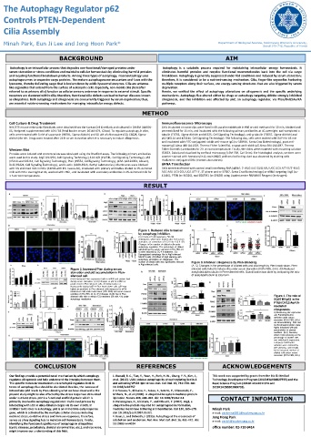
Figure 1. Reduced cilia formation
by autophagy inhibition.
(A) Effects of 5 nM rapamycin, 50 mM
chloroquine, which were treated after 24 h serum
starvation, on conversion of LC3-I into LC3-II. (B)
Changes in the number of ciliated cells under
autophagy regulation. The percentage of ciliated
cells(induced by serum starvation-0.5% FBS, 24
h) were reduced by CQ 4 h treatment. (C)
Dysregulated autophagy flux in Atg5-silenced
NIH/3T3 cells. (D) Effect of Atg5 silencing with
autophagy stimulation on ciliogenesis. The
number of ciliated cells was significantly reduced Figure 3. Inhibited ciliogenesis by Pten-silencing.
in Atg5-silenced cells. (A–C) Changes in the percentage of ciliated cells and cilia length by Pten knock-down. Pten-
Figure 2. Increased Pten during serum silenced cells failed to induce cilia under serum starvation (0.5% FBS, 24 h). (D) Reduced
starvation and p62 accumulation in Pten- acetylated alpha tubulin in Pten-silenced cells. Quantification was done by comparing the ratio
silenced cell. of acetylated tubulin to total form.
(A,B) Increased Pten expression both in mRNA and protein level
during serum starvation. (C,D) Changes of p62 at mRNA or
protein level in Pten-silenced cells. (E) Observation of
fluorescently-labeled p62 in Pten knock-down cells. p62 was
highly accumulated in Pten-silenced cells compared to siCtrl-
transfected cells under both basal (10% FBS) and serum starved
condition (0.5% FBS, 24 h). (F) Changes of p62 flux in Pten-
silenced cells with or without CQ treatment (50 mM, 4 h) under Figure 4. The role of
autophagy modulation. SQSTM1/p62 in the
PTEN/DVL2/AurKA-
mediated
ciliogenesis.
A) Monitoring p62 expression
and Pten/Dvl2/AurKA
activities under serum
starvation (0.5% FBS, 24 h)
via immunoblotting. Dvl2 and
AurKA phosphorylation were
highly enhanced with p62
accumulation in starved
condition. (B,C) Recovery
effect of p62-silencing on the
percentage of ciliated cells
and Dvl2/AurKA expression.
Enhanced Dvl2/AurKA
activities were restored by
p62-silencing, and it finally
led to increased number of
ciliated cells under serum
starvation (0.5% FBS, 24 h).
CONCLUSION REFERENCES ACKNOWLEDGEMENTS
Our findings provide a potential novel mechanism by which autophagy 1. Russell, R. C., Tian, Y., Yuan, H., Park, H. W., Chang, Y. Y., Kim, J., This work was supported by grants from the Bio & Medical
regulates ciliogenesis and help understand the interplay between them. et al. (2013). ULK1 induces autophagy by phosphorylating Beclin-1 Technology Development Program (2015M3A9B6027555) and the
The specific molecular mechanisms via which p62 regulates Dvl2 in and activating VPS34 lipid kinase. Nat. Cell Biol. 15, 741–750. doi: Basic Science Program (2016R1A5A1011974 and
terms of autophagy flux should be elucidated. Besides, the increase of 10.1038/ncb2757 2019R1A2B5B03069738).
intracellular p62 levels by Pten-silencing what we have observed in the 2. Ichimura, Y., Kirisako, T., Takao, T., Satomi, Y., Shimonishi, Y.,
present study might be also affected by the stress responses stimulated Ishihara, N., et al. (2000). A ubiquitin-like system mediates protein
under nutrient stress. p62 is a functional scaffold protein which is lipidation. Nature 408, 488–492. doi: 10.1038/35044114 CONTACT INFOMATION
primarily involved in autophagy regulation in multi-level process by 3. Nakatogawa, H., Ichimura, Y., and Ohsumi, Y. (2007). Atg8, a
interacting with LC3 or ubiquitinated cargos via its own motifs. In ubiquitin-like protein required for autophagosome formation,
addition to its roles in autophagy, p62 is an immediate-early response mediates membrane tethering and hemifusion. Cell 130, 165–178. Minah Park
gene, which is activated by the multiple cellular stresses including doi: 10.1016/j.cell.2007.05.021 e-mail: parkmina0821@sookmyung.ac.kr
nutrient stress, oxidative stress and immune responses, therefore, 4. Kaur, J., and Debnath, J. (2015). Autophagy at the crossroads of Jong Hoog Park
serves as a key signaling hub to maintain cell homeostasis. Finally, catabolism and anabolism. Nat. Rev. Mol. Cell. Biol. 16, 461–472. doi: e-mail: parkjh@sookmyung.ac.kr
identifying the functional significance of autophagy in ciliopathies 10.1038/nrm4024
(cystic diseases, polydactyly, skeletal abnormalities, etc.), and vice versa, office number: 02-710-9414
might improve our understanding of this field.