
Page 17 - H. Cell signaling
P. 17
HDM-induced chemokine CCL20 regulates Chronic Inflammation and
Remodeling of Airway in Mouse Model of Bronchial Asthma
Shin-Young Park , Min-Jeong Kang , Yun Young Lee , So Young Lee , and Joong-Soo Han 1,2
1
2
2
2
1 Biomedical Research Institute and Department of Biochemistry & Molecular Biology, College of Medicine,
2 Department of Biomedical Sciences, Graduate School for Biomedical Science & Engineering, Hanyang University, Seoul, Republic of Korea
BACKGROUND AIM
* Chronic inflammatory response in the airway in asthma
- Increased Th2 lymphocytes, eosinophils HDM extracts were shown to induce C-C chemokine ligand 20 (CCL20) secretion in airway epithelial cells
- Activated mast cells for the recruitment of immature dendritic cells (DCs) to the lung 4 . CCL20 has been found to be
upregulated by a variety of inflammatory cytokines and it plays an important role in innate immunity 5 .
Moreover, CCL20 was reported to be upregulated in inflammatory diseases, such as allergic airway
disease, rheumatoid arthritis . Recent work showed that HDM-induced CCL20 was related to oxidative
stress and early allergic airway responses 3 . However, the precise role of CCL20 induced by HDM in the
allergic airway inflammation remains to be elucidated.
Thus, the purpose of this study is investigated the mechanism by which HDM induces C-C chemokine
Molecular Therapy - Nucleic Acids 2020 191000-1014 ligand 20 (CCL20) expressions to promote chronic inflammation and remodeling of airway in mouse
model of bronchial asthma. This work further illuminates the mechanism of bronchial asthma and
* House dust mites (HDM) are one of the most common sources of allergens associated with symptomatic provide a new therapeutic approaches for lung fibrosis.
allergic airway diseases. HDM components and airway epithelial cell receptors that interact to mediate allergic
sensitization, airway inflammation and airway remodeling
METHODS
Generation of the HDM-induced asthma model
Eight-week-old BALB/c mice were sensitized by intranasal administration of HDM (30 ug/mouse) or saline (Sham) twice a week for 3 weeks. For CCL20 neutralizing study, mice were sensitized by intranasal administration of CCL20 (0.5 ug/mouse) or saline
(Sham) 5 times a week for 4 weeks. Mice were treated with anti-CCL20 (5 ug/mouse) intranasally every time CCL20 was administered. The mice were killed, and the deliver efficiency of the proteins and the pathological changes in the lungs were analyzed.
Airway hyper-responsiveness (AHR) analysis
Mice were anesthetized (pentobarbital sodium, intraperitoneally), ventilated (flexiVent 5.1®; SCIREQ, Montreal, Canada) and challenged with a saline aerosol followed by increasing concentrations of methacholine (MeCh; Sigma-Aldrich). Aerosols were
generated with an ultrasonic nebulizer (Omron Healthcare, Kyoto) and delivered to the inspiratory line of the flexiVent using a bias flow of medical air. Measurements were made twice at 1min intervals following each concentration of MeCh aerosol.
Bronchoalveolar lavage (BAL) fluid analysis
The lungs were lavaged with 1 mL Hank’s balanced salt solution (HBSS) via the tracheostomy tube. Total cell numbers were counted with a hemocytometer. After the procedure, BAL fluid was centrifuged, and then smears of BAL cells were prepared by
cytocentrifugation (Cytospin3, Thermo, Billerica, MA) at 1,000 rpm for 3 min. BAL cells were stained with Hemacolor Staining Kit (Merck Millipore, Darmstadt) counted, and classified as neutrophils, eosinophils, lymphocytes, or macrophages. The cells were
differentially counted until the total counted number reached at least 200, using standard hemocytologic procedures to count macrophages and eosinophils.
RESULTS
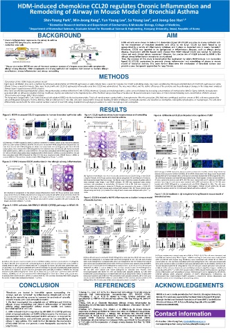
Figure1. HDM increased CCL20 expression in human bronchial epithelial cells Figure 4. CCL20 regulates airway hyper-responsiveness and remodeling Figure 6. HDM-induced CCL20 participatesin the regulation of EMT
of airway in mouse model of bronchial asthma.
(A) Heatmap of mRNA expression levels in asthma compared to healthy control using microarray datasets
(GEO accession number GSE4302). (B) BEAS-2B cells were treated with HDM (10ug/ml) for indicated time. (C)
Cells were treated with HDM (10ug/ml) for 30min and stained with anti-CCL20 (green). (D) Cells in 96-well
culture plates were treated with HDM (10ug/ml) for the indicated times (E) BEAS-2B cells were transfected
with 100nM Dentin-1 siRNAs or control siRNA for 72hr and then stimulated with HDM (10ug/ml) for 30 min.
(F) Cells on 96-well culture plates were transfected with 100nM Dectin-1 siRNAs or control siRNA for 72h and
then stimulated with HDM (10ug/ml) for 24hr (G) Cells were transfected with 100nM Dectin-1 siRNAs or
control siRNA for 72hr and then stimulated with HDM (10ug/ml) for 30 min. *. p <0.05.
Figure 2. HDM induces CCL20 production in the allergic airway inflammation.
(A) Heatmap of mRNA expression levels in asthma compared to healthy control using microarray
datasets (GEO accession number GSE4302). (B-D) mRNA and proteins from lung of each mouse were
(A) Eight-week-old BALB/c mice were intranasally administratered CCL20 0.25ug or Anti-CCL20 analyzed with qPCR and western blot. (E-H) Beas-2B cells were treated with HDM (10ug/ml) for 24 hr.
2.5ug for 4 weeks. (B-D) Tissue sections were stained using H&E, PAS and Masson’s Trichrome The cells were harvested, and total RNA was isolated using TRIzol reagent. Proteins (E) and mRNAs (F,
and observed by bright-filed microscopy. The fibrotic area (C) and the number of goblet cells (D) G) were analyzed by western and qPCR. (H) Cells were stained with anti-E-cadherin. (I) mRNA and
were analyzed using Image J and SABIA software. (E) AHR was measured using airway proteins from lung of each mouse were analyzed with qPCR and western blot. (L-O) Cells were
resistance and compliance to metacholine. (F) Immune Cells in BAL fluid were counted. A treated with CCL20 (100ng/ml) or co-treated with anti-CCL20 (10ug/ml) for 24hr. The cells were
representative of each group is shown (n=5). Results are presented as the mean ± S.E.M. (G) harvested, and total RNA was isolated using TRIzol reagent. Proteins (L) and mRNAs (M, N) were
Proteins from lung of each mouse were analyzed with western blot. (H) Tissue sections were analyzed by western and qPCR. (O) Cells were stained with anti-E-cadherin. *. p <0.05.
stained using anti-IL-1β. (I, K) mRNAs from lung of each mouse were analyzed with qPCR. *. p
(A) Schedule for preparation of the allergic airway inflammation model. (B) Tissue sections were stained <0.05. Figure 7. CCL20-mediated IL-1β is required for lung fibrosis in mouse model of
using anti-CCL20. (C, D) mRNA and proteins from lungs were analyzed with qPCR and western blot. (E) CCL20
was measured from homogenates of the lung of each mouse. (F) Tissue sections were stained using H&E, Figure 5. CCL20 is related to NLRP3 inflammasome activation in mouse model bronchial asthma
PAS and Masson’s Trichrome. (G, H) The fibrotic area and the number of goblet cells were analyzed using of bronchial asthma.
Image J and SABIA software. *. p <0.05.
Figure 3. HDM activates Akt-ERK1/2-GSK3β-C/EBPβ pathways in BEAS-2B
cells.
(A) Tissue sections were stained using anti-α-SMA or TGF-β1. (B, C) The cells were harvested, and
(A) Beas-2B cells were treated with CCL20 (100ng/ml) for indicated time (B-D) Cells were treated total RNA was isolated using TRIzol reagent. mRNAs from lung of each mouse were analyzed with
with CCL20 (100ng/ml) or co-treated with anti-CCL20 (10ug/ml) for 4hr. (E) Cells were treated qPCR. (D) IMR-90 cells (human lung fibroblasts) were transfected with 100nM NF-κB siRNAs or
(A, B) Beas-2B cells were transfected with vector or C/EBP β for 48hr, and then treated with Der f 2 (10ug/ml) with CCL20 (100ng/ml) or co-treated with anti-CCL20 (10ug/ml) for 6hr (F) Proteins from lung of control siRNA for 72hr and then stimulated with IL-1β (100ng/ml) for 24hr. Cells were stained with
for 30min. (B) Cells were stained with anti-CCL20 (green). (C, D) Cells were transfected with control siRNA or each mouse were analyzed with western blot. (H) Tissue sections were stained using anti-NLRP3. anti-F-actin and anti-α-SMA. (E-H) IMR-90 cells were pretreated with TGF-β1 inhibitor, SB431542, for
C/EBPβ siRNA for 48hr, and then treated with Der f 2 (10ug/ml) for 30min. (D) Cells were stained with anti- (H) Cells were treated with CCL20 (100ng/ml) for indicated time. (I, J) Cells were transfected with 1hr and stimulated with TGF-β1 (50ug/ml) for 24 hr. (E) Cells were stained with anti-F-actin and anti-
CCL20 (green). (E, F) Cells were pretreated with 1uM GSK-inhibitor for 1hr and stimulated with HDM (10ug/ml) 100nM NF-κB siRNAs or control siRNA for 72hr and then stimulated with CCL20 (100ng/ml) for α-SMA. (F-H) The cells were harvested, and total RNA was isolated using TRIzol reagent. mRNA levels
for 15min (E) or 30min (F). (G, H) Cells were pretreated with 2uM ERK1/2 inhibitor, PD98059, for 1hr and 6hr. (K) Cells were treated with CCL20 (100ng/ml) for indicated time. Proteins were were analyzed by qPCR. *. p <0.05. (I) Proposed model of HDM-induced CCL20 production signaling in
stimulated with HDM (10ug/ml) for 15min (G) or 30min (H). (I, J) Cells were pretreated with 1uM AKT immunoprecipitate with NLRP3 for 24hr and blotted with ubiquitin antibody. (L) Cells were chronic inflammation and remodeling of airway. HDM-induced CCL20 is regulated by Akt-ERK1/2-
inhibitor, LY294002, for 1hr and stimulated with HDM (10ug/ml) for 15min (I) or 30min (J (K, L) Cells were treated with CCL20 (100ng/ml) or co-treated with anti-CCL20 (10ug/ml) for 30min. Proteins were C/EBP1β pathway which enhanced activation of NLRP3 inflammasome. Furthermore, we
transfected with 100nM Dectin-1 siRNAs or control siRNA for 72hr and then stimulated with HDM (10ug/ml) immunoprecipitated with NLRP3 for 24hr and blotted with ubiquitin antibody. (M-O) Cells were demonstrated that CCL20-mediated NLRP3 activation is required for chronic inflammation and
for 15 min. Proteins were analyzed by western. *. p <0.05. transfected with 100nM NLRP3 siRNAs or control siRNA for 72hr and then stimulated with CCL20 profibrotic process in the remodeling of airway in lung asthma.
(100ng/ml) for 6hr. Proteins and mRNAs were analyzed by western and qPCR. *. p <0.05.
CONCLUSION REFERENCES ACKNOWLEDGEMENTS
Fibroblasts are located in interstitial spaces surrounding the 1.Huang, F.L., Liao, E.C. & Yu, S.J. House dust mite allergy: Its innate immune
airways, and are “activated” by inflammatory stimuli such as IL-1β response and immunotherapy. Immunobiology 223, 300-302 (2018). IMR90 cells were kindly provided by Prof. Kim E.G. (ChungbukUniversity,
during the remodeling process to increase the synthesis of cytosolic 2.Porsbjerg, C. et al. IL-33 is related to innate immune activation and Korea). This work was supported by the Basic Science Research Program
sensitization to HDM in mild steroid-free asthma. Clin Exp Allergy 46, 564-574
smooth muscle actin and extracellular matrix . (2016). through the National Research Foundation of Korea (NRF), funded by the
1. We first investigated the potential role of HDM-induced CCL20 in 3.Hong, G.H. et al. Clusterin Modulates Allergic Airway Inflammation by Ministry of Science, ICT, & Future Planning, Republic of Korea (NRF-
allergic airway remodeling and chronic inflammation of bronchial Attenuating CCL20-Mediated Dendritic Cell Recruitment. J Immunol 196, 2021- 2018R1A1A1A05022185).
asthma using experimental asthma mouse models and human 2030(2016).
bronchial epithelial cell lines. 4.Nathan, A.T., Peterson, E.A., Chakir, J. & Wills-Karp, M. Innate immune
2. HDM-induced CCL20 is regulated by Akt-ERK1/2-C/EBP1β pathway responses of airway epithelium to house dust mite are mediated through beta-
glucan-dependent pathways. J Allergy Clin Immunol 123, 612-618 (2009).
which enhanced activation of NLRP3 inflammasome. Furthermore, we 5.Schutyser, E., Struyf, S. & Van Damme, J. The CC chemokine CCL20 and its Contact information
demonstrated that CCL20-mediated NLRP3 activation is required for receptor CCR6. Cytokine Growth Factor Rev 14, 409-426 (2003).
chronic inflammation and profibrotic process in the remodeling of 6. Weckmann, M. et al. Critical link between TRAIL and CCL20 for the activation
airway in lung asthma. This work further illuminates the mechanism of TH2 cells and the expression of allergic airway disease. Nat Med 13, 1308- -First author : Shin-Young Park, ttokttoki@hanyang.ac.kr
of bronchial asthma and provide a new therapeutic approaches for 1315 (2007). -Corresponding author: Joong-Soo han, jshan@hanyang.ac.kr
lung fibrosis.