
Page 15 - H. Cell signaling
P. 15
p62 enhances the auto-ubiquitination of TRAF6 induced by lipopolysaccharides
Sung-Hwan Cho, Kyung-Hye Roh and Eui-Ju Choi
Laboratory of Cell Death and Human Diseases, Department of Life Sciences, Korea University, Seoul 02841, South Korea
BACKGROUND AIM
In order to better understand the mechanism by which p62 enhances the
≫Toll-like receptor 4(TLR4) signaling pathway TRAF6 activity, we investigated a role of p62 in the oligomerization and auto-
Lipopolysaccharides(LPS) triggers molecular rearrangement of a receptor complex and oligomerization of TLR4.[1] Upon TLR4 oligomerization, TLR4 ubiquitination of TRAF6.
recruits its downstream adaptor proteins through the Toll-interleukin-1 receptor (TIR) domain, which is an intracellular signaling domain found in myeloid
differentiation primary response gene 88 (MyD88), interleukin-1 receptors, Toll receptors. There are four downstream adaptors containing TIR domain: TIR METHODS
domain-containing adaptor protein (TIRAP), MyD88, TIR domain-containing adaptor inducing IFN-β (TRIF), TRIF-related adaptor molecule (TRAM). [2]
TLR4 uses different combinations of adaptors to determine downstream signaling: TIRAP/MyD88, TRIF/TRAM. The signal is divided into MyD88
dependent pathway and MyD88 independent pathway (TRIF dependent pathway). The MyD88 dependent pathway is responsible for expression of >>Ubiquitination assay
proinflammatory cytokines, while the MyD88 independent pathway mediate induction of type Ⅰ interferons. MyD88 has a death domain (DD) in addition to For cell-based assay of ubiquitination in MEF cells, the cells were treated for 15min with LPS.
the TIR domain. MyD88 recruits other DD containing molecules, IL-1R-associated kinase (IRAK) family kinases, through homotypic interactions. Upon Cells were lysed in buffer A [1% Triton X-100(v/v), 5mM EGTA, 20mM Tris-HCl (pH 7.4), 150mM
TLR4 activation, activation of IRAK family kinases activates TNF receptor-associated factor 6 (TRAF6).[3] Recruitment/activation of TRAF6, along with sodium chloride, 0.5% sodium deoxycholate, 12mM β-glycerophosphate, 10mM sodium fluoride,
other E2 ubiquitin protein ligases, activates a complex containing TGF-β-activated kinase 1 (TAK1), TAK1-binding protein 1 (TAB1), TAB2.[4] TAK1 then 1mM phenylmethyl sulfonyl fluoride, 2μg/ml aprotinin, and 2μg/ml leupeptin] containing 5mM N-
activates downstream IKK (inhibitor of κ light chain gene enhancer in B cells kinase) pathway. A complex containing IKKα , IKKβ and IKKγ phosphorylates ethylmaleimide. The lysates were incubated for 10min at 50℃ and then subjected to
inhibitor of κ light chain gene enhancer in B cells (IκB) proteins. This phosphorylation of IκB proteins leads to the ubiquitination of IκB proteins and then immunoprecipitation with antibody to TRAF6. The resulting precipitates were visualized by
immunoblot analysis with antibody to ubiquitin.
degradation of IκB proteins. A transcription factor NF- κB can be translocated to nucleus through degradation of IκB proteins. This translocation controls the
expression of proinflammatory cytokines. [5] >>Co-immunoprecipitation
≫TRAF6 Cells were lysed in NETN lysis buffer [0.5% Nonidet P-40 (v/v), 1 mM EDTA (pH 8.0), 50 mM
TRAF6 is a E3 ubiquitin ligase that mediates polyubiquitination on substrate via its N-terminal RING finger domain and an adaptor protein that mediates a Tris-HCl (pH 8.0), 120 mM sodium chloride, 1mM dithiothreitol, 0.2mM sodium orthovanadate,
wide array of protein-protein interaction through its C-terminal TRAF domain. As TRAF family [6], TRAF6 contains three domains: a N-terminal RING 10mM sodium fluoride, 1 mM phenylmethyl sulfonyl fluoride, 2 μg/ml aprotinin, and 2 μg/ml
leupeptin]. Cell lysates were incubated at 4 °C for 16 h with appropriate antibodies and then for
domain (residues 70-106), a ZINC finger domain (150-259) and a C-terminal TRAF domain (299-530). TRAF family proteins are divided according to the an additional 1 hour in the presence of protein G-coupled Sepharose beads (Amersham
type of interacting protein through its C-terminal TRAF domain. Ubiquitination functions as a multi-use signal mark because substrates can be modified by Biosciences). The resulting precipitates were washed twice with cell lysis buffer and boiled for 5
the addition of ubiquitin molecules to lysine residues of proteins. Ubiquitination of target proteins involves a three-step enzymatic process. The unique E1, min after which samples were resolved by SDS sample buffer.
ubiquitin-activating, enzyme activates ubiquitin by binding it to one of its own cysteine. It then passes ubiquitin onto E2, ubiquitin-conjugating, enzyme
through its own cysteine. E3, ubiquitin ligase, enzyme transfers ubiquitin to its final protein target. TRAF6 catalyzes the synthesis of unique polyubiquitin >>In situ proximity ligation assay
chains, Lys63-linked polyubiquitination. These Lys63-linked chains are important for downstream activation of IKK by recruiting and activating TAK1.[6] The assay was performed with the use of a Duolink Ⅱ fluorescence kit(Olink Bioscience)
Oligomerization and auto-ubiquitination of TRAF6 are often used as a read-out for TRAF6 activation.
≫p62 (sequestosome1) >>In vitro binding assay
p62 is a scaffold protein that possesses PB1 dimerization domain and TRAF binding sequence, and UBA ubiquitin-associating domain. Recent studies
HEK293T cells were transfected with Flag-TRAF6 for 48 h, and the cells were lysed in buffer A.
have shown that p62 plays a role in cell signaling by influencing TRAF6 activity. However, its functional role in inflammatory signaling is controversial. Cell lysates were subjected to immunoprecipitation with anti-TRAF6 antibody and incubated for
Kwon J et al. found that p62 negatively regulated TLR4 signaling through functional regulation of the TRAF6-ECSIT complex.[7] In the contrary, NF-κB an additional 1 hour with 20μl of protein G-coupled Sepharose beads. The resulting precipitates
activation mediated by CD40, a member of the tumor necrosis factor (TNF) receptor family, was positively regulated by TRAF6/p62 complex.[8] Similarly, were washed twice with cell lysis buffer. The immunopellets were incubated with MBP-tagged
Moscat J et al. reported that p62 facilitates Lys63-linked polyubiquitination of TRAF6 and thereby mediates nerve growth factor-induced activation of the protein for 1 hour at 4℃ in a binding buffer [50mM Tris-HCl (pH 7.5), 150mM sodium chloride,
NF-κB pathway. [9] 2mM EDTA, 1mM dithiothreitol, 0.1% Nonidet P-40 (v/v), 0.1mM sodium orthovanadate, bovine
serum albumin 5μg/μl]. Bead-bound proteins were washed twice with washing buffer [50mM
HEPES, 150mM sodium chloride, 1mM EDTA, 1mM dithiothreitol, 0.1% tween-20(v/v)] and
boiled for 5min after which samples were resolved by SDS sample buffer.
RESULTS
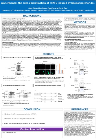
p62 promotes the LPS-induced ubiquitination of TRAF6 siRNA-mediated depletion of p62 in TRAF6 -/- MEFs
mitigates the oligomerization of TRAF6 induced by LPS B
Figure 1. Genetic ablation of p62 impairs the LPS-induced TRAF6 C
Figure 3. siRNA-mediated knockdown of p62 in TRAF6 -/- MEFs
auto-ubiquitination in MEFs alleviates the oligomerization of TRAF6 upon LPS stimulation.
MEF cells were treated for 15min with LPS, and then subjected ubiquitination assay. Indicated expression vectors were transfected in TRAF6 -/- MEFs. The cells were then
subjected to an in situ proximity ligation assay with PLA probes, according to the
manufacturer’s protocol. More than 50 cells were examined in experiment and the protein–
protein interaction was quantified from one experiment.
p62 increases the oligomerization of TRAF6 Identification of the p62 domains that bind to TRAF6
A B A
Figure 4. The UBA and PB1 domains of p62 are important for
the interaction between p62 and TRAF6.
(A) Various mutant p62 constructs : p62 full length (1-440), p62 PB1 (1-124),
p62 ΔPB1 (125-440), p62 ΔUBA (1-385), p62 ΔPB1ΔUBA (125-385). (B), (C) In
vitro binding assay was performed.
Figure 2. p62 promotes the oligomerization of TRAF6, while it
does not affect the TRAF6-Ubc13 interaction
(A),(B) HEK293T cells were transfected with indicated expression vectors then
subjected co-immunoprecipitation .
CONCLUSION REFERENCES
1. Visintin A, Iliev DB, Monks BG, Halmen KA, Golenbock DT. MD-2.
>> p62 induces the LPS-induced auto-ubiquitination of TRAF6. Immunobiology. 2006;211(6-8):437‐447. doi:10.1016/j.imbio.2006.05.010
2. O’Neill LA, Bowie AG. The family of five: TIR-domain-containing adaptors in
Toll-like receptor signalling. Nat Rev Immunol 2007;7:353–64.
3. Deguine J, Barton GM. MyD88: a central player in innate immune signaling.
F1000Prime Rep. 2014;6:97. Published 2014 Nov 4. doi:10.12703/P6-97
>> p62 promotes the LPS-induced oligomerization of TRAF6. 4. Chen ZJ. Ubiquitin signalling in the NF-kappaB pathway. Nat Cell Biol.
2005;7(8):758‐765. doi:10.1038/ncb0805-758
5. Lu YC, Yeh WC, Ohashi PS. LPS/TLR4 signal transduction pathway.
Cytokine. 2008;42(2):145‐151. doi:10.1016/j.cyto.2008.01.006
>>The PB1 and UBA domains of p62 are crucial for the TRAF6-p62 interaction. 6. Walsh MC, Lee J, Choi Y. Tumor necrosis factor receptor- associated factor
6 (TRAF6) regulation of development, function, and homeostasis of the
immune system. Immunol Rev. 2015;266(1):72‐92. doi:10.1111/imr.12302
7. Kim MJ, Min Y, Kwon J, et al. p62 Negatively Regulates TLR4 Signaling via
Functional Regulation of the TRAF6-ECSIT Complex. Immune Netw.
Contact information 2019;19(3):e16. Published 2019 Jun 12. doi:10.4110/in.2019.19.e16
8. Seibold K, Ehrenschwender M. p62 regulates CD40-mediated NFκB
activation in macrophages through interaction with TRAF6. Biochem Biophys
Res Commun. 2015;464(1):330‐335. doi:10.1016/j.bbrc.2015.06.153
E-mail : izakpine@gmail.com 9. Wooten MW, Geetha T, Seibenhener ML, Babu JR, Diaz-Meco MT, Moscat
J. The p62 scaffold regulates nerve growth factor-induced NF-kappaB
activation by influencing TRAF6 polyubiquitination. J Biol Chem.
2005;280(42):35625‐35629. doi:10.1074/jbc.C500237200