
Page 19 - H. Cell signaling
P. 19
Nitration of protein phosphatase 2A increases via Epac1/PLCε/CaMKⅡ/HDAC5/iNOS
cascade during cAMP-induced decidualization of human endometrial stromal cells
So Young Lee, Yun Young Lee, Jung-Sub Choi, Kyeong Soo Kim, Do Sik Min, Shin-Young Park*, Joong-Soo Han*
Biomedical Research Institute and Department of Biochemistry and Molecular Biology, College of Medicine, Hanyang University,
222 Wangsimni-ro, Seongdong-gu Seoul 04763, Republic of Korea, E-mail : jshan@hanyang.ac.kr, ttokttoki@hanyang.ac.kr.
Abstract Introduction
Decidualization effects on hESCs to grow constantly and mature for successful for embryo
Decidualiation of endometrial stroma is an essential differentiation process for embryo implantation implantation and maintenance of pregnancy during menstrual cycle [1]. Decidualization
and maintaining pregnancy. We previously reported that protein phosphatase 2A (PP2A) acts as a requires elevated progesterone and intracellular cAMP levels [2,3]. PP2A in involved many
key mediator during cAMP-induced decidualization of human endometrial stromal cells (hESCs). cellular processes but was not known until now to play any role in the decidualization of
However, its activation mechanism has been remained veiled in these models. In present study, hESCs. However, mechanisms to regulates the distribution of PP2A activation during cAMP-
we aimed to reveal the mechanism how to induce the nitration of PP2A catalytic subunit (PP2Ac) induced decidualization are not understood. Post-translational modification of PP2Ac such as
during cAMP-induced decidualization of hESCs. Taken together, our results suggest that the phosphorylation, methylation or nitration regulates PP2A activity. We therefore propose that
PP2Ac nitration during cAMP-induced decidualization of hESCs is induced through the Epac1- Epac1-Rap1-PLCε signaling cascade promotes CaMKⅡ phosphorylation, leading to
Rap1-PLCε-CaMKⅡ-HDAC5-iNOS signaling pathway. upregulation of iNOS protein expression through inactivation of HDAC5, which in turn is
responsible for nitration in PP2A during cAMP-induced decidualization of hESCs.
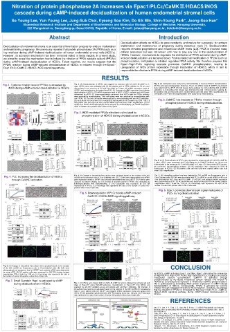
RESULTS
Fig. 1. Tyrosine nitration level of PP2Ac is increased by Fig. 1. (A) Decidualization of hESCs was triggered by treatment with 0.5 mM 8-Br-cAMP for 3 Fig. 2. (A) Cell lysates were analyzed by immunoblotting to measure HDAC5 phosphorylation.
(B) The cells were photographed. Scale bar, 40 µm. (C) mRNA levels of IGFBP1 and prolactin
days. hESCs were also pretreated with 30 nM okadaic acid (a PP2A inhibitor) for 30 min and
iNOS during cAMP-induced decidualization in hESCs. differentiated in the presence of 0.5 mM 8-Br-cAMP for 3 days. (B) mRNA expression levels of were determined by qPCR. (D) Cell lysates were analyzed by immunoblotting with anti-iNOS
IGFBP1 and prolactin were determined by qPCR. (C) Heatmap of mRNA expression levels during antibodies. (E) Intracellular NO release was determined by DAF-FM fluorescence staining. (F)
decidualization in hESCs. (D) mRNA expression level of iNOS, IGFBP1, prolactin and eNOS were NO-positive cells and total cells were counted within each field under 100× magnification. (G)
determined by qPCR. (E) Immunoprecipitation performed from hESCs with the PP2Ac antibody Cell lysates and PP2Ac immunoprecipitates were analyzed by immunoblotting.
were analyzed by immunoblotting. Total cell lysates were also analyzed by immunoblotting with
anti-PP2Ac. (F) The total cell lysates were analyzed by immunoblotting with anti-iNOS antibodies. Fig. 3. CaMKⅡ has impacts on PP2Ac nitration through
(G) Intracellular NO levels were then evaluated by staining with an NO-sensitive dye (DAF-FM). (H)
NO-positive cells and total cells were counted within each field under 100× magnification. (I) Cell phosphorylation of HDAC5 during decidualization.
lysates and PP2Ac immunoprecipitates were analyzed by immunoblotting. (K) mRNA expression
levels of IGFBP1 and prolactin were measured by qPCR.
Fig. 2. iNOS-mediated PP2Ac nitration is stimulated by
phosphorylation of HDAC5 during decidualization in hESCs.
Fig. 3. (A, B) Decidualization of hESCs was stimulated by treatment with 0.5 mM 8-Br-cAMP for
3 days. hESCs were pretreated with 5 µM Bapta-am (a CaMKⅡ inhibitor) for 30 min and
differentiated in the presence of 0.5 mM 8-Br-cAMP for 3 days. (C) Using whole hESC lysates,
we performed immunoblotting analyses. (D) NO production was measured by DAF-FM
fluorescence in hESCs. (E) NO-positive cells and total cells were counted within each field
under 100× magnification.
Fig. 4. PLC increases the decidualization of hESCs Fig. 4. (A) Changes in intracellular free calcium were calculated based on the variation of the 340 Fig. 5. (A) Intracellular calcium level was detected by 340 nm/380 nm fluorescence ratio in
nm/380 nm fluorescence ratio in Fura-2-labeled cells. (B, C) Cells were photographed and mRNA
Fura-2-labeled cells. (B) Cells were transfected with PLC ε siRNA or control siRNA for 48 h and
through CaMKⅡ activation. gene expression levels of IGFBP1 and prolactin were determined using qPCR. (D) Proteins were then differentiated for 3 days with 8-Br-cAMP. (C) IGFBP1 and prolactin mRNA expression
analyzed by western blotting. (E) The amount of nitrated tyrosine in PP2Ac was measured by were determined by qPCR. (E) NO production was analyzed through DAF-FM staining (original
immunoprecipitation with immunoblotting. (F) NO production was measured by DAF-FM magnification 200×). Scale bar, 100 µm. (F) Percentage ratio represents the ratio of the
fluorescence in hESCs. (G) Percentage ratio represents the ratio of the number of control NO- number of control NO-positive cells to that of total cells.
positive cells to that of total cells.
Fig. 6. Epac1 promotes downstream signal molecules of
Fig. 5. Downregulation of PLCε blocks cAMP-induced PLCε during decidualization.
CaMKⅡ-HDAC5-iNOS signaling pathway.
Fig. 6. (A) Changes in intracellular free calcium were calculated based on the variation CONCLUSION
of the 340 nm/380 nm fluorescence ratio in Fura-2-labeled cells. (B) Cells were
photographed and expression level of IGFBP1 and prolactin mRNA were determined
by using qPCR. (E) NO-positive cells were measured by DAF-FM staining (original
magnification 200×). Scale bar, 100 µm. (F) Percentage ratio represents the ratio of In hESCs, cAMP activates Epac1, and then, Epac1 stimulates the conversion
the number of control NO-positive cells to that of total cells. of Rap1-GDP to Rap1-GTP. Active Rap1 activates PLCε, leading to increased
intracellular calcium concentration. CaMKⅡ is activated through
Fig. 7. Small G protein Rap1 is activated by cAMP phosphorylation by IP 3 -related intracellular calcium release. The function of
HDAC5 as a transcriptional repressor is inhibited by CaMKⅡ phosphorylation.
during decidualization in hESCs. Fig. 7. (A) hESCs were treated with 0.5 mM 8-Br-cAMP for 50 min, followed by cell lysis and pull- NO is synthesized by increasing iNOS protein expression in cAMP-induced
down of Rap-GTP using RalGDS-Sepharose. Quantification of Rap1-GTP from hESCs was
decidualization of hESCs. Consequently, PP2Ac is activated by tyrosine
analyzed by anti-GST pulldown assay and probing with anti-Rap1 antibodies. (B) Changes in nitration through increased intracellular NO production. Therefore, we propose
intracellular free calcium are calculated based on the variation of the 340 nm/380 nm fluorescence that the nitration of PP2Ac is increased by the Epac1-Rap1-PLCε -CaMKⅡ-
ratio in Fura-2-labeled cells. (D) IGFBP1 and prolactin mRNA expression levels were analyzed by HDAC5-iNOS signaling pathway during cAMP-induced decidualization of
qPCR. (F) NO production was detected by DAF-FM staining (scale bar, 100 µm). (G) Percentage hESCs.
ratio represents the ratio of the number of control NO-positive cells to that of total cells.
REFERENCES
Lee, S. Y., Lee, Y. Y., Choi, J. S., Yoon, M. S. & Han, J. S. (2016) Phosphatidic acid induces
decidualization by stimulating Akt-PP2A binding in human endometrial stromal cells. Febs. J.
283, 4163–4175.
Yoon, M. S., Koo, J. B., Jeong, Y. G., Kim, Y. S., Lee, J. H., Yun, H. J., Lee, K. S. & Han, J. S.
(2007) Phospholipase D1 as a key enzyme for decidualization in human endometrial stromal
cells, Biology of reproduction. 76, 250-8.
Ohama, T., & Brautigan, D. L. (2010). Endotoxin conditioning induces VCP/p97-mediated and
inducible nitric-oxide synthase-dependent Tyr284 nitration in protein phosphatase 2A. Journal of
Biological Chemistry, 285(12), 8711-8718.
Millward., T. A., Zolnierowicz., S. & Hemmings., B. A. (1999) Regulation of protein kinase
cascades by protein phosphatase 2A, Elsevier Science. 24.