
Page 29 - G. Cell differentiation. division. and death
P. 29
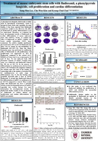
ABSTRACT RESULTS RESULTS
A phenylpyrrole fungicide, fludioxonil, is widely (A)
used in agricultural environment, which is 1.0
present in many fruits and vegetables. In some DMSO
studies, it has been reported that fludioxonil 0.8 10-7 M
caused low weight in rats. However, the effect 10-5 M
of fludioxonil on cardiac differentiation is not Beating cardiomyocytes (%) 0.6
yet understood. Therefore, we evaluated the 0.4
early developmental toxicity of fludioxonil on
cardiac differentiation of mouse embryonic 0.2
stem cells (mESCs). Firstly, to confirm the 0.0
effect of fludioxonil on mESCs viability, the 1 3 5 8 11 14 17 20 23 26 29 32 35 38 42
water soluble tetrazolium (WST) assay Days after seeding
conducted. The mESCs viability significantly
decreased under 50% at 10-5 M fludioxonil, but Figure 4. Effect of fludioxonil on mESCs-derived
there was no change in cell morphology by (B) cardiomyocytes.
fludioxonil (10-5-10-9 M). Then, the colony Fludioxonil After EB formation, EBs were collected, picked at day 3
formation assay was performed to confirm the and seeded at 5 EBs / well. EBs were cultured in 0.1%
effect of fludioxonil on cell proliferation. Cell lolk gelatin-coated 6-well plates using differentiation medium
*
proliferation was suppressed by 10-5 M 80 Day 5 until the contraction stops. Contracting cells were
fludioxonil, compared to the control (0.1% 60 Day 10 observed by microscope (IX73, Olympus).
Day 15
DMSO) at 5 days, but it was re-increased at 10
and 15 days. To test embryoid body (EB) Colony formation (% of control) 40
formation capacity of mESCs, hanging drop 20 SUMMARY
assay was conducted and fludioxonil reduced
the EB size at 10-5 M. In the process of 0 DMSO 10 -7 10 -5
differentiation to cardiomyocytes derived from Concentration (M)
mESCs, 10-5 M fludioxonil completely inhibited
the beating ratio (the ratio of the number of Figure 2. Effect of fludioxonil on mESCs
contracting cells to the number of attached EBs) proliferation.
mESCs were seeded at 50 cells / well using
of cardiomyocytes at early stage of undifferentiation medium containing LIF and treated with
differentiation (day 5), but the beating ratio −7 −5 M). After
gradually increased after 5 days at 10-5M 0.1% DMSO and fludioxonil (10 and 10
culture by date (5, 10 and 15 day), mESCs were stained
fludioxonil. It seemed that fludioxonil delayed with crystal violet solution. (A) mESCs was pictured by
the differentiation of mESCs to cardiomyocytes date. (B) Increase of proliferation of mESCs was
at 10-5 M compared to control. These results estimated and quantified by image J program. CONCLUSION
imply that fludioxonil may have a potential
toxicity on the developmental process of mESCs, (A)
especially into cardiac lineage. For more In this study, it was confirmed that
information for developmental toxicity of fludioxonil inhibited the proliferation of
fludioxonil, further studies on the mechanisms mESC and inhibited the myocardial
of fludioxonil to induce altered cell differentiation process.
proliferation and cardiac differentiation of Additionally, further studies on the
mESCs are needed. mechanism of fludioxonil are needed to
identify potential toxicity to the
RESULTS developmental toxicity of fludioxonil.
Fludioxonil REFERENCES
(B)
150 1. Kang HY, Choi YK, Jeung EB, et al.:
Advanced developmental toxicity test method
Cell proliferation (% as control) 100 * 2. Nuoya Yin, Shengxian Liang, Shaojun Liang,
based on embryoid body’s area. Reproductive
Toxicology 72: 74-85, 2017.
et al.: DEP and DBP induce cytotoxicity in
50
mouse embryonic stem cells and abnormally
Environmental Pollution 236: 21-32, 2018.
0 -9 -8 -7 -6 -5 enhance neural ectoderm development.
DMSO 10 10 10 10 10 3. Tui Neri, Valeria Merico, Maurizio Zuccotti,
Concentration (M) et al.: The differentiation of cardiomyocytes
Figure 1. Effect of fludioxonil on mESCs from mouse embryonic stem cells is altered by
proliferation. Figure 3. Effect of fludioxonil on mEB formation. dioxin. Toxicology Letters 202: 226-236, 2011.
mESCs were seeded at 5× 10 3 cells / well using mESCs were seeded at 800 cells / 25 μl / droplet using
undifferentiation medium containing LIF in 96-well plates differentiation medium without LIF and treated with
and exposed to various concentrations of fludioxonil 0.1% DMSO and fludioxonil (10 - 10 −5 M). About 64
−9
−9
(10 - 10 −5 M) for 5 days. The cell proliferation was droplets were seeded on the lid of Petri dishes for 3 days.
measured by EZ-Cytox at 540 nm. Data were presented as And then, (A) EB formation was pictured by microscope
the means ± SD of triple experiments. *p<0.05 compared (IX73, Olympus). (B) Reduction of EB size was
with control, 0.1% DMSO. estimated and quantified by image J program.