
Page 17 - G. Cell differentiation. division. and death
P. 17
Regulation of death receptor-mediated apoptosis via
USP8 post-translation modification of c-FLIP L
Chi Hyun Hwang¹ , Manhyung Jeong¹ , Eun-Woo Lee², Daehyeon Seong¹, Jinho Seo³, Jaewhan Song¹
,#
,#
¹Department of Biochemistry, College of Life science and Biotechnology, Yonsei University, Seoul 120-749, Korea ²Metabolic Regulation Research Center, Korea Research Institute of Bioscience
and Biotechnology (KRIBB), Daejeon 34141, Korea ³Environmental Disease Research Center, Korea Research Institute of Bioscience and Biotechnology (KRIBB), Daejeon 34141, Korea
BACKGROUND & AIM
Apoptosis can be induced by various stimuli including anti-Fas, TRAIL and TNFα induced activation of death receptors. Cellular FLICE-
like inhibitory protein (c-FLIP) has been identified as a protease-dead, procaspase-8-like regulator of death ligand-induced apoptosis
based on observations that c-FLIP impedes death ligand-induced apoptosis by binding to FADD and/or caspase-8 or -10 in a ligand-
dependent fashion.c-FLIP is a family of alternatively spliced variants that primarily exists in human cells as long (c-FLIP L ) and short (c-
FLIP s ) splice variants. Here, we demonstrated that ubiquitin-specific peptidase 8 (USP8), a c-FLIP L deubiquitinase, has a regulatory
mechanism for c-FLIP L de-ubiquitination and protein level up-regulation. USP8 depletion accelerates death ligand-induced extrinsic
apoptosis due to FLIP L destabilization. Additionaly, we found a correlation between elevated c-FLIP L protein levels and melanoma and
cervical cancers, due to up-regulation of USP8 mRNA and protein levels. Therefore, we used the ME-180 cervical cancer xenograft model
to show that USP8 depletion attenuated tumor growth upon TRAIL injection. These finding indicate that pharmacological regulation of
USP8 may provide an effective strategy to treat various cancers.
RESULTS & METHODS
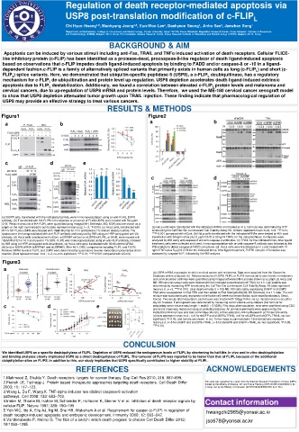
Figure1 Figure2
a b a b
c d e
c d
(a) 293FT cells, transfected with the indicated plasmids, were immunoprecipitated using an anti-FLAG, USP8
antibody. (b) Transfected with HA-FLIPL in the absence or presence of FLAG-USP8, were treated with 50 μg/ml
CHX. Relative amounts of HA-FLIPL were quantified using ImageJ(NIH, Bethesda, MD, USA) and are shown as a
graph on the right (normalized to actin) (dots represent mean±s.d.; n =3, *P‹0.05). (c) HeLa cells, transfected with (a) HeLa cells were transfected with the indicated siRNAs and treated as in a. Cell loss was determined by ATP
WT or MT FLAG-USP8, were treated with 10μM MG132 for 10 h and lysed in 1% sodium dodecyl sulfate. The levels using the CellTiter-Glo Luminescent Cell Viability Assay Kit. All bars represent mean ±s.d.; n=3, **P‹0.01,
lysates were immunoprecipitated with anti-FLIP antibody and analyzed by WB using an HRP-conjugated anti-Ub ***P‹0.001 compared with siCont. (b) HeLa cells transfected with the indicated siRNAs were treated at 48 h post-
antibody. (d) HeLa cells ransfected with siCont, siUSP8#1+siCont or siUSP8+siFLIPL, at 30nM, were treated with transfection with 50ng/ml αFas plus 5 μg/ml CHX or 50ng/ml TRAIL for the indicated times. Cell lysates were
10μM MG132 for 10 h and lysed in 1% SDS. FLIPL was immunoprecipitated using an anti-FLIP antibody, followed analyzed by WB using anti-caspase-8 and anti-caspase-3 antibodies. (c) TRAIL for the indicated times. After ligand
by WB using an HRP-conjugated anti-Ub antibody. (e) HeLa cells were transfected with 30nM control siRNA treatment, cells were collected and lysed. Immunoprecipitation with an anti-caspase-8 antibody was followed by the
(siCont) or USP8 siRNA (siUSP8#1 and siUSP8#2). After 48 h, DISC components including FLIPL and FLIPS. WB analysis to detect caspase-8-FADD complexes. (d) HeLa cells were transfected as in c and treated with 10
Relative mRNA levels of FLIPL and USP8 were determined by quantitative reverse transcription-polymerase chain ng/ml TNFα plus 5 μg/ml CHX for the indicated times. After ligand treatment, TNFR1 complex II formation was
reaction.(Bars represent mean ±s.d.; n=3, ns=non-significant, **P 0.01, ***P‹0.001 compared with siCont). assessed by caspase-8 IP, followed by the WB analysis
Figure3
a b
(a) USP8 mRNA expression levels in cervical cancer and melanoma. Data were acquired from the Oncomine
Database and re-analyzed. (b) Relative amounts of USP8, FLIPL or FLIPS normalized to actin levels in melanoma
and cervical cancer cell lines were quantified using ImageJ software (NIH) and are shown as a graph.(c) HeLa and
ME-180 cervical cancer cells were treated with the indicated concentrations of TRAIL for 4 h. Cell viability was
determined by measuring ATP levels using the CellTiter-Glo Luminescent Cell Viability Assay Kit (dots represent
mean±s.d.; n =3, ***P‹0.001). (d-e) Approximately 2 × 10⁶ ME-180 cells stably expressing shGFP or shUSP8
RNA were resuspended in 50μl PBS and then added to 50μl Matrigel(354234; BD Sciences) in a 1:1 ratio. The cell
mixture was subcutaneously implanted in the flanks of 6-week-old female Balb/c nude mice(Narabiotech, Seoul,
Korea). Two weeks after inoculation, each mouse was treated with 150μg TRAIL via i.p. injection once every other
day for 3 weeks. Tumor growth was determined by measuring tumor volume using calipers (the formula for
c d e calculating tumor volume was length × width2 × 0.5236). Fifty days afterinoculation, mice were sacrificed using CO2,
and tumor mass was measured using an analytical balance. All animal experiments were approved by the
Institutional Animal Care and Use Committee (IACUC) of the Laboratory Animal Research at Yonsei University.
(d) dots represent mean ±s.d.; n=5 for shGFP and shUSP8+TRAIL, n=6 for shUSP8 and shGFP+TRAIL, ns=non-
significant, *P‹0.05, and tumors are shown in (e). Tumor mass is summarized in f (horizontal bars represent
mean±s.d.; n=5 for shGFP and shUSP8+TRAIL, n=6 for shUSP8 and shGFP+TRAIL,ns=non-significant, *P‹0.05,
**P‹0.01).
CONCULSION
We identified USP8 as a specific deubiquitylase of FLIPL. Depletion of USP8 reduced the endogenous levels of FLIPL by shortening its half-life. In vivo and in vitro deubiquitylation
and binding analyses clearly implicated USP8 as a direct deubiquitylase of FLIPL. The turnover of FLIPS was reported to be faster than that of FLIPL because of the additional
ubiquitylation sites on FLIPS. In addition to this, our study implicates that USP8 specifically contributes to higher stability of FLIPL.
REFERENCES ACKNOWLEDGEMENTS
1.Mahmood Z, Shukla Y. Death receptors: targets for cancer therapy. Exp Cell Res 2010; 316: 887–899.
2.French LE, Tschopp J. Protein-based therapeutic approaches targeting death receptors. Cell Death Differ This work was supported by a grant from the National Research Foundation of Korea (NRF),
funded by the Ministry of Science, ICT and Future Planning (NRF-2015R1A3A2066581) (to
2003; 10: 117–123. J Song) and by the Ministry of Education (2012R1A6A3A04040105) (to E-WL).
3.Wang L, Du F, Wang X. TNF-alpha induces two distinct caspase-8 activation
pathways. Cell 2008; 133: 693–703.
4.Irmler M, Thome M, Hahne M, Schneider P, Hofmann K, Steiner V et al. Inhibition of death receptor signals by Contact information
cellular FLIP. Nature 1997; 388: 190–195
5.Yeh WC, Itie A, Elia AJ, Ng M, Shu HB, Wakeham A et al. Requirement for casper (c-FLIP) in regulation of hwangchi2985@yonsei.ac.kr
death receptor-induced apoptosis and embryonic development. Immunity 2000; 12: 633–642.
6.Vandenabeele P, Melino G. The flick of a switch: which death program to choose Cell Death Differ 2012; jso678@yonsei.ac.kr
19:1093–1095.