Page 21 - G. Cell differentiation. division. and death
P. 21

Establishment of canine primary myoblast culture


                                                                                                                                                       and myotubes differentiation


                                                                                                               Feriel Yasmine Mahiddine  , Jin Wook Kim, Min Jung Kim                                                                                                                                                                                                         a,*
                                                                                                                                                                                                                                     a


                   a    Department of Theriogenology and Biotechnologies, College of Veterinary Medicine, Seoul National University, South Korea.


                                                                                                                                                                       *   Corresponding author: tinia19@snu.ac.kr


        INTR ODUC TION                                                                                                                                                                                                                                  R E S ULTS


       Skeletal                                      muscle                                     cells                          originates                                              from                             their


       precursors, satellite cells or also called myogenic


       stem cells, and are multinucleated and form striated


       tissues. The use of skeletal muscle cells in vitro


       became common in aging or muscle pathology


       research; however, most studies use established cell


       lines rather than primary isolated cells, which can


       provide results closer to the in vivo state. In this


       study, we tried to establish a protocol for myoblast


       isolation, culture and differentiation in canine


       species.


        MATE R IALS  AND ME THODS


        1. Animal use


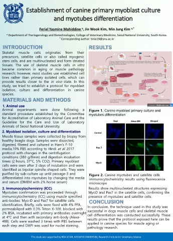
       Animal                                    experiments                                                     were                             done                             following                                           a                 Figure 1. Canine myoblast primary culture and


       standard procedure established by the Committee                                                                                                                                                                                                   myotubes differentiation


       for Accreditation of Laboratory Animal Care and the


       Guideline for the Care and Use of Laboratory


       Animals of Seoul National University.


       2. Myoblast isolation, culture and differentiation


       Muscle tissue samples were collected by biopsy from


       healthy beagle dogs. Samples were dissected,


       digested, filtered and cultured in Ham’s F-10


       media,10% FBS according to Hindi et al. 2017


       protocol with changes in the centrifugation


       conditions (380 g/8min) and digestion incubation


       times (2 hours, 37°C, 5% CO2). Primary myoblast


       cells were seen after 3 days of incubation and were


       identified as bipolar spindle-shaped cells. They were


       purified by sub-culture up until passage-3 and                                                                                                                                                                                                    Figure 2. Canine myotubes and satellite cells


       differentiated into myotubes by changing the media                                                                                                                                                                                                immunocytochemistry results using fluorescence


       and serum (DMEM with 2% horse serum)                                                                                                                                                                                                              microscope


       3. Immunocytochemistry (ICC)                                                                                                                                                                                                                    Results show multinucleated structures expressing


       Myotubes confirmation was proceeded through                                                                                                                                                                                                     MyoD and Pax7 in the satellite cells, confirming the


       immunocytochemistry (ICC) using muscle specified                                                                                                                                                                                                presence of myotubes and satellite cells.


       anti-bodies: Myo-D and Pax7 for satellite cells                                                                                                                                                                                                   C ONC LUS ION


       identification. Briefly, cells were fixed with 4% PFA,                                                                                                                                                                                        In conclusion, the technique used in this study was


       permeabilized with 0.3% Triton X-100, blocked with                                                                                                                                                                                            successful in dogs muscle cells and skeletal muscle


       2% BSA, incubated with primary antibodies overnight                                                                                                                                                                                           cell differentiation was conducted successfully. These


       at 4°C and then with secondary anti-body (Alexa-                                                                                                                                                                                             results prove that the protocol exposed here can be


       488). Cells were washed 3 times with PBS between                                                                                                                                                                                              applied in canine species for muscle aging or


       each step and DAPI was used for nuclei staining.

                                                                                                                                                                                                                                                     pathology research.


                                                                                        This study was supported by RDA (CCAR, #2018R1C1B6009536), Research Institute for Veterinary Science.
   16   17   18   19   20   21   22   23   24   25   26