
Page 51 - F. Cell biology
P. 51
Cell Death according to Silica Nanoparticles manufactured by different methods
Baek Jin Ee, Shin Jae Hoon, Lee Jong Seong
Institute of Occupation & Environment, KCOMWEL, Incheon, Republic of korea
BACKGROUND AIM
Silica nanoparticles can be roughly divided into two types: wet route base The purpose of this study, was to determine
synthesis method and thermal route base (Pyrogenic) synthesis method. the difference in cytotoxicity and cell death
Differences in the silica production method may affect the physicochemical mechanism according to the physicochemical
properties of silica nanoparticles, and may cause different cytotoxicity properties of silica nanoparticles by confirming
results for silica nanoparticles. However, there have been few studies on the the difference in cytotoxicity according to the
cytotoxicity of silica nanoparticles made by other methods. method of manufacturing silica nanoparticles.
METHODS
4 types of Silica nanoparticles (Table 1) dispersed in a culture Table 1. Silica Nanoparticles (SiNPs)
solution containing 10% FBS were diluted to a concentration of 0- Method Category Properties Size [nm] Manufacturer
5000 ug/ml, and then put into A549 cells and reacted for 24 hours. Sigma Aldrich
Colloidal Hydrophilic 20 - 30
The size and stability of the dispersed silica nanoparticles were Wet - base (Ludox Ⓡ TM)
analyzed using an electron microscope and a zeta analyzer. CCK-8 Mesoporous Hydrophilic 200 Sigma Aldrich
reagent was used to confirm proliferation, and LDH assay was Fumed (602) Hydrophilic 20 - 30 HW nanomaterial
pyrogenic
performed. And the ROS generation of the cells was confirmed.
Fumed (606) Hydrophobic 20 - 30 HW nanomaterial
RESULTS
The viability of cells exposed to silica nanoparticles (colloidal silica and porous silica) made by wet pretreatment and cells
exposed to fumed silica nanoparticles showed a difference, and LDH secretion and ROS generation due to cell damage also
showed different results.
Table 2. Hydrodynamic properties of silica nanoparticles
Hydrodynamic polydispersity
Silica ζ-potential [mV]
diameter [nm] index (PDI)
Colloidal SiNPs 154 0.383 -9.09
Mesoporous SiNPs 326 0.125 -8.71
Fumed (602) SiNPs 309 0.471 -8.94
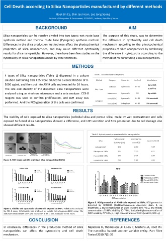
Figure 1. TEM image and XRD analysis of Silica nanoparticles (SiNPs) Fumed (606) SiNPs 230 0.161 -8.64
A549
3
Colloidal SiNPs
Mesoporouse SiNPs
2 Fumed (602) SiNPs
Relative ROS generation Fumed (606) SiNPs
1
0
0 1 2 3 4 0 1 2 3 4 0 1 2 3 4 0 1 2 3 4
SiNPs concentration
Figure 3. ROS generation of A549 cells exposed to SiNPs. ROS generation
detected by DCFH-DA (Dichlorofluorescein diacetate) stain. 0, no
Figure 2. viability and cytotoxicity of A549 cells exposed to SiNPs. Viability was analyzed treatment; 1, low concentration of SiNPs (viability 80% ↑); 2, low-middle
by CCK-8 assay. Cytotoxicity was analyzed by Lactate Dehydrogenase(LDH) assay. The concentration of SiNPs (viability 80~70%); 3,middle-High concentration of
cells were treated with SiNPs and incubation in 37 ℃ CO 2 incubator for 24 hours. SiNPs viability 70~50%,; 4, High concentration of SiNPs (viability 50% ↓)
CONCLUSION REFERENCES
In conclusion, differences in the production method of silica Napierska D, Thomassen LC, Lison D, Martens JA, Hoet PH.
nanoparticles can affect the cytotoxicity and cell death The nanosilica hazard: another variable entity. Part Fibre
mechanism. Toxicol 2010;7(1):39