
Page 53 - F. Cell biology
P. 53
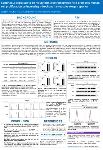
Continuous exposure to 60 Hz uniform electromagnetic field promotes human
  cell proliferation by increasing mitochondrial reactive oxygen species


                                       2
             1
                          2
                                                     2
  Donghwa Suh , Dae Yong Um , Sang Hyeon Im , Gwan Soo Park , Kiwon Song 1,*
                   BACKGROUND                                                  AIM
    Electromagnetic waves are generated when an electric field and a magnetic field
                                                           A considerable amount of wire is distributed on the ground and
   change with time, and can be transmitted without a medium substance.  underground of the city to maintain modern civilization, and the majority of
   Electromagnetic spectrum can be divided into ionizing radiation and non-ionizing
   radiation whether carrying sufficient energy to ionize other molecules and atoms or  them generate extremely low frequency electromagnetic fields (ELF-EMF).
                                                          Thus we are continuously exposed to ELF-EMF in daily life. However, the
   not. One of the non-ionizing radiation ranges, radiofrequency electromagnetic
   fields (RF-EMF), had classified as ‘possibly carcinogen to humans (Group B)’ in  physiological effects of the ELF-EMF and their mechanisms are not clearly
                                                          documented. Previous studies showed controversial results depending on the
   2011. WHO/International Agency for Research on Cancer (IARC) announced that  frequency and conditions of ELF-EMF, and types of cells: cells can either
   they can increase the risks of brain cancer. The extremely low-frequency  proliferate or undergo apoptosis, DNA damage with an increase or decrease
   electromagnetic fields (ELF-EMF), another non-ionizing radiation, also have been  in the level of ROS. In this study, we aim to monitor the cellular effect of
   suggested for physiological effects, but their generalized risks have not been
   systemically studied. Here, we investigate the cellular effects of ELF-EMF and their  strong EMFs, greater than 10 mT, on various human cells. Also, we
                                                          investigate the specific molecular mechanisms of ELF-EMF for cellular effects.
   specific molecular mechanisms.
                                                METHODS
   We designed a closed-type device to generate uniform ELF-EMF. We focused the sole effect of ELF-EMF excluding thermal effects by installing the digitalized
   thermometer. In this device, 6 dishes can be simultaneously exposed to ELF-EMF with an intensity of 10 to 16 mT. Cells were plated in 35 mm culture plates, and
   exposed to a 60 Hz uniform ELF-EMF over 10mT after 18 h of incubation. We tested the cellular effect of these ELF-EMF on various human cells, including human
   cervical carcinoma HeLa, liver cancer stem cells Huh7 and Hep3B, immortalized normal hepatic cell MIHA, and normal fibroblast IMR90. Both MTT assay and cell
   counting system were conducted to monitor cell proliferation. To observed changes in protein expression following ELF-EMF treatment, we normalized band intensity
   after western blot analysis. Flow cytometer analysis was performed for cell cycle analysis with propidium iodide (PI) staining, intracellular and mitochondrial ROS
   levels were measured with H2-DCFDA and MitoSOX staining, respectively.
                                                RESULTS

                    EMF strength (mT)
    Cell type
               10       14        16
     HeLa   119.54±3.45*  138.60±3.03*** 142.28±12.78***
     Huh7  119.12±1.90*** 127.29±4.43*** 128.45±2.82***
     Hep3B  106.77±2.52*** 108.43±1.92***  109.20±3.24**
                                                                        Figure 2. Continuous exposure to a uniform ELF-
     MIHA   119.56±2.52**  119.54±0.94**  116.97±5.55*                  EMF decreases the expression of the tumor
                                                                        suppressor p16 in HeLa
     IMR90  115.47±3.99***  109.14±1.81**  102.42±2.11 ns

   Table 1. A continuous exposure to 60 Hz ELF-EMF
   promotes cell proliferation in various human cells
   HeLa, Huh7, Hep3B, MIHA and IMR90 were continuously  Figure 1. Cell proliferation by a continuous
   exposed to 10, 14, 16 mT ELF-EMF for 72 h.  exposure to 14 mT is reversible and
   Values of *P<0.05, **P<0.01, ***p<0.001 were considered  transient
   statistically significant,  and P>0.05  was  considered  After 6 h of ELF-EMF exposure, each cells
   statistically not significant.        were further incubated without exposure of
                                         ELF-EMF for up to 72 h.
                                                                        Figure 3. Continuous exposure to a uniform ELF-
                                                                        EMFs increases phosphorylation of Erk1/2 in
                                                  Figure 4. Continuous exposure  HeLa
                                                  to a uniform ELF-EMF does
                                                  not disturb cell cycle
                                                  HeLa was exposed to an ELF-
                                                  EMF at 14 mT for up to 72 h.
                                                  Cell cycle distributions of HeLa
                                                  exposed to the ELF-EMFs were
                                                  determined by flow cytometry
                                                  using PI staining.

             CONCLUSION                        REFERENCES
   •  A continuous exposure to ELF-EMF accelerated the  •  Song K, Im SH, Yoon YJ, Kim HM, Lee HJ,
     proliferation in HeLa, Huh7, Hep3B, MIHA, and IMR90  et al. (2018) PLOS ONE 13(7): e0199753.  Figure 5. Continuous exposure to a uniform ELF-
     cells.                                •  Lekovic, M.H., Drekovic, N.E., Granica, N.D.  EMF increases intracellular and mitochondrial
   •  The increased cell proliferation by ELF-EMF was  et al. (2020) Environ Sci Pollut Res.  ROS in HeLa
     transient and reversible.                https://doi.org/10.1007/s11356-020-10039-0  Contact information
   •  ELF-EMF decreased the expression of the tumor  ACKNOWLEDGEMENTS
     suppressor p16, and increased the expression of                    1 Department of Biochemistry, College of Life Science
     phosphorylated Erk1/2.                                             and Biotechnology, Yonsei University, Seoul, 03722,
   •  ELF-EMF activated cell cycle, but did not lead to  This work was supported by the ICT R&D  South Korea
     significant cell cycle perturbation.  program of MSIT/IITP. [2019-0-00102, A  2 Department  of  Electrical  Engineering,  Pusan
   •  ELF-EMF triggered slight increase of intracellular and  Study on Public Health and Safety in a  National University, Pusan, 46241, South Korea
     mitochondrial ROS levels, suggesting that ELF-EMF  Complex EMF Environment]
     may activate the electron transport system.                        althehdghk@yonsei.ac.kr & bc5012@yonsei.ac.kr 1,*
                                                                                        1