
Page 63 - D. Cancer biology
P. 63
Differential sensitivity of BRAF- and RAS-mutated cells to
autophagy inhibition
Hojin Yeom , Sung-Hee Hwang , Byeal-I Han , and Michael Lee 1,2,*
1
2
1
1 Division of Life Sciences, College of Life Sciences and Bioengineering, Incheon National University, Incheon 22012, Republic of Korea
2 Institute for New Drug Development, Incheon National University, Incheon 22012, Korea
Abstract Introduction
The deregulation of autophagy is frequent in malignancy. In this study, we investigated the therapeutic effects of autophagy on tumor cells Autophagy
containing BRAF-mutations, which have been found to be present in up to 70% of all melanomas. Sensitivity to BRAF inhibitor PLX4720 is well A crucial process involved in cell
correlated with BRAF mutational status in melanoma cell lines. Dose response analyses showed that all the BRAF mutant melanoma cell strains homeostasis and cell survival by
were highly sensitive to PLX4720 compared to cells with WT BRAF. We observed the increased basal autophagic flux in BRAF-mutated cells removing unnecessary proteins and
damaged organelles.
compared with WT BRAF cells as determined by LC3 conversion and immunofluorescence using the tandem probe RFP-GFP-LC3. Combination An important regulator of survival /
treatment with PLX4720 and early autophagy inhibitor SBI-0206965 resulted in significantly greater cytotoxicity than PLX4720 or SBI-0206965 death response by chemotherapy in
treatment alone regardless of BRAF mutation status. However, late autophagy inhibitor hydroxychloroquine did not show synergistic effect in melanoma.
combinations with PLX4720. We further investigated changes in PLX4720 sensitivity of cells with an ATG5 gene-knockout (KO) in BRAF-
mutated A375P cells and SK-MEL-2 cells bearing WT BRAF. We found that the ATG5 KO led to PLX4720 resistance in both cell lines. We also The tandem probe RFP-GFP-LC3
observed synergistic anti-survival effects of MEK inhibitor trametinib when combined with PLX4720 in SK-MEL-2 cells but not in A375P cells. This method can confirm autophagic
Taken together, our results suggest that while combined inhibition of early autophagy with BRAF inhibitor might be a viable therapeutic avenue for flux because it shows GFP and RFP
BRAF mutant melanomas, targeted therapies that inhibit MEK signaling may prove a more effective treatment strategy for BRAF wild-type signals before fusion with lysosomes
melanomas. (This work was supported by the National Research Foundation of Korea (NRF) grant funded by the Ministry of Science and ICT and only RFP signals after fusion
(MIST) (NRF-2019R1F1A1048733)). with lysosomes.
Results
Fig. 1 Growth response to PLX4720 in cell lines with differing BRAF mutational status. (A) Whole cell lysates were
prepared from four cell lines with differing BRAF mutational status. Melanoma cells are BRAF homozygous or
heterozygous for wild-type (W), or V600E (E) mutation. The phosphorylated forms of MEK and ERK were detected
by immunoblotting using anti-phospho-MEK and anti-phospho-ERK antibodies. The same blots were reprobed with
anti-MEK and anti-ERK antibodies to confirm the similar expression level of MEK and ERK proteins in all lanes. (B)
A total of four melanoma cell lines were also treated with PLX4720 from 0.1 to 10 μM for 72 h. Cell growth was then
evaluated with WST-1 assay. The relative viability of cells treated with vehicle alone was regarded as 100%. Values
represent the mean ± SD of quadruplicate determinants from one of three representative experiments.
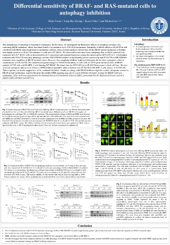
Fig. 2. PLX4720-induced autophagy in cell lines with differing BRAF mutational status. (A)
Basal autophagy was evaluated in four cell lines with differing BRAF mutational status by
monitoring LC3 conversion by western blotting. (B) Autophagic flux was also measured using
the ptfLC3 plasmid that simultaneously expresses mRFP- and GFP-tagged LC3 protein. Ratio
quantification of autophagosomal LC3 puncta to autopahgolysosomal LC3 puncta (n = 20). (C)
Representative images of fluorescence microscopy analysis of yellow- and red-fluorescence
EGFP-LC3B punctate vesicular structures, indicative of autophagosomes and autolysosomes
Fig. 3. Combination effect of PLX4720 and autophagy inhibitor on cell survival. (A) A total of four cell lines accumulation, respectively. The cells were transfected with the ptfLC3 plasmid and treated
were also treated with PLX4720 from 0.1 to 20 μM or with early autophagy inhibitor SBI-0206965 (10 μM) for with 1 μM PLX4720 or with 10 μM HCQ for 24 h alone or in combination. Early
72 h alone or in combination. (B) Melanoma cell lines were treated with PLX4720 from 0.1 to 20 μM or with autophagosomes show GFP-mRFP colocalization whereas late, acidic autophagosomes (arrows)
late autophagy inhibitor HCQ (10 μM) for 72 h alone or in combination. In (A) and (B), cell growth was then lose GFP and appear red. Scale bars, 10 μm. Lower panel: Ratio quantification of
evaluated with WST-1 assay. The relative viability of cells treated with vehicle alone was regarded as 100%. autophagosomal LC3 puncta to autopahgolysosomal LC3 puncta (n = 10).
Values represent the mean ± SD of quadruplicate determinants from one of three representative experiments.
Fig. 4. Combination effect on cell survival of PLX4720 and MEK
inhibitor in parental and ATG5 KO cells. (A) and (B) A375P (A),
SK-MEL-2 (B), and their ATG5 KO counterparts were treated
with increasing concentrations of PLX4720 ranging from 0.1 to
200 μM or with MEK inhibitor trametinib (0.1 μM) for 72 h alone
or in combination. Cell growth was then evaluated with WST-1
assay. The relative viability of cells treated with vehicle alone was
regarded as 100%. Values represent the mean ± SD of
quadruplicate determinants from one of three representative
experiments. (C) and (D) Cell lysates were prepared from A375P,
SK-MEL-2, and their ATG5 KO counterparts treated with the
indicated concentrations of 20 μM PLX4720 for 24 h. The
phosphorylated forms of MEK (A) and ERK (B) were detected by
immunoblotting using anti-phospho-MEK and anti-phospho-ERK
antibodies. The same blots were reprobed with anti-MEK and
anti-ERK antibodies to confirm the similar expression level of
MEK and ERK proteins in all lanes.
Conclusion
The Combination treatment with PLX4720 and early autophagy inhibitor SBI-0206965 resulted in significantly greater cytotoxicity than each mono-treatment regardless of BRAF mutation status.
The ATG5 KO led to PLX4720 resistance in both cell lines.
MEK inhibitor trametinib treatment combined with PLX4720 was synergistic anti-survival effects in SK-MEL-2.
Our results suggest that while combined inhibition of early autophagy with BRAF inhibitor might be a viable therapeutic avenue for BRAF mutant melanomas, targeted therapies that inhibit MEK signaling may prove
a more effective treatment strategy for BRAF wild-type melanomas.