
Page 39 - D. Cancer biology
P. 39
Novel Interaction of MLK3/AURKC as a Therapeutic Target in Breast Cancer
Jin Young Min 1, 2 , Eun Hee Han 1, *
¹Division of Bioconvergence Analysis, Korea Basic Science Institute (KBSI), Cheongju 28119, Republic of Korea,
²College of Pharmacy, Chungnam National University, Daejeon 34134, Republic of Korea
ABSTRACT METHOD
Aurora C kinase (AURKC) has an activity with tumorigenesis in breast
cancer and may be a relevant cancer target. AURKC is an interesting
target for the development of anticancer therapy, but its signaling
network has not been fully characterized. Here we report the
identification of MLK3 as one of the AURKC binding partners and
abnormal interaction of MLK3-AURKC induce tumorigenic activity.
MLK3 was screened as one of the AURKC binding partners through
the CUPID assay. In addition, expression and interaction of
AURKC/MLK3 were highly observed in breast cancer cells compare to
breast normal cells. Furthermore, correlation with novel interaction
and tumorigenesis was confirmed by synergistic anti-tumor effects of
combination treatment of pan-MLKs inhibitor (CEP1347) and AURKB/C
inhibitor (GSK1070916) in MDA-MB-231 cells. Therefore, complex of
AURKC/MLK3 can be a new anti-cancer therapeutic target in breast
cancer and provides a clue to understand concealed role of AURKC in
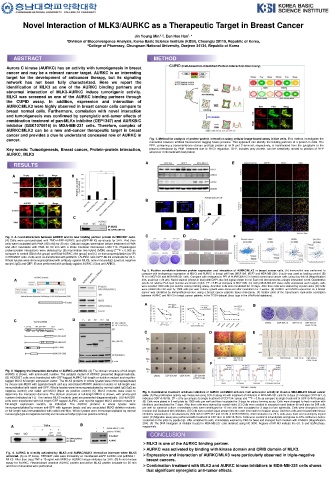
cancer. Fig. 1. Method for analysis of protein-protein interaction using cellular image-based assay in live cells. This method investigates the
interaction between artificial fluorescence tagging fusion proteins. This approach can identify the binding partners of a protein in cells. The
RFP, containing a transmembrane domain and bait protein at its N and C-terminal, respectively, is translocated from the cytoplasm to the
Key words: Tumorigenesis, Breast cancer, Protein-protein interaction, plasma membrane by PMA treatment due to PKCδ regulation. GFP, includes prey protein, can be selectively moved to position of RFP
whenever it interacts with bait protein.
AURKC, MLK3
RESULTS A E
A B
B
C
Fig. 2. A novel interaction between AURKC and its new binding partner protein in HEK293T cells. C D F
(A) Cells were co-transfected with TMD-mRFP-AURKC and eGFP-MLK3 constructs for 24 h. And then
cells were incubated with PMA (500 nM) for 20 min. Cellular images were taken before treatment of PMA
and after incubation with PMA for 20 min with a Zeiss Confocal microscope LSM 710. Physiological
protein-protein interactions were detected by (B) mammalian two hybrid (M2H) assay [***P < 0.005 as
compare to control (Bind+Act group) and Bind-AURKC+Act group] and (C) co-immunoprecipitation (co-IP)
in HEK293T cells. Cells were co-transfected with pcDNA3.1-AURKC and eGFP-MLK3 constructs for 24 h.
Whole lysates were immunoprecipitated with antibody against MLK3, nomal mouse IgG (used as negative
control; IgG) and GFP. IB was performed with antibody against AURKC (Goat anti-ARK3).
Fig. 5. Positive correlation between protein expression and interaction of AURKC/MLK3 in breast cancer cells. (A) Immunoblot was performed to
compare with endogenous expression of MLK3 and AURKC in breast cell lines (MCF10A, MCF7 and MDA-MB-231). β-actin was used as loading control. (B)
A B PLA in MCF10A and MDA-MB-231 cells. Compare with endogenous PPI of AURKC/MLK3 in breast normal and cancer cells using duo-link kit (Magnification
40X, scale bar = 20 μm). Nuclei stained (shown in blue) with DAPI, duo-link signal shown in red. Each red dot represents a single interaction event. Quantitative
results for relative PLA spot number are shown (right). [*P < 0.05 as compare to MCF10A]. (C) Using MDA-MB-231 were stably expressed each targets, cells
were seeded 1000 cells per well for colony forming assay. And then cells were incubated for 10 days. After then cells were stained by crystal violet. (D) Cells
were plated into soft agar by 1000 cells per well. Colony growth were observed after incubation for 4 weeks. (E) AURKC and MLK3 expression on a breast
cancer was identified by immunofluorescence using a breast cancer patients tissue microarray. (F) Scatter plots of the Spearman’s rank-order correlation
between AURKC and MLK3 in breast cancer patients in the TCGA dataset (brca_tcga in the cBioPortal database).
A B
C D
C D E
F
Fig. 3. Mapping the interaction domains of AURKC and MLK3. (A) The domain structure of full-length
AURKC is shown, with amino-acid number. The catalytic mutant of AURKC presented diagrammatically.
(B) HEK293T cells were transfected with GFP-tagged AURKC full-length or deletion mutant and myc/his-
tagged MLK3 full-length expression vector. The MLK3 proteins in whole lysates were immunoprecipitated
by mouse anti-MLK3 with agarose beads and any associated AURKC deletion mutants or full-length was
immunoblotted with rabbit anti-GFP. Whole lysates were immunoprecipitated by normal rabbit IgG (IgG as
negative control) and rabbit anti-MLK3 (Input as positive control) (C) MLK3 mutants were used to Fig. 6. Combination treatment of kinase inhibitors of AURKC and MLK3 enhance their anti-cancer activity in invasive MDA-MB-231 breast cancer
determine the interaction domains. The domain structure of full-length MLK3 is shown, with amino-acid cells. (A)The proliferative activity was measured using CCK-8 assay kit with treatment of inhibitors in MDA-MB-231 cells for 8 days (C indicates CEP1347, G
number (indicated as FL). The various MLK3 mutants used are presented diagrammatically. (D) HEK293T indicates GSK1070916). [*P < 0.05 as compare to single treatment of CEP1347 group and ***P < 0.05 as compare to single treatment of GSK1070916 group].
cells were transfected with full-length GFP-tagged AURKC and myc/his-tagged MLK3 deletion mutant or (B) Cells were plated in 6 well plate by 1000 cells per well and then incubated for 3 days for colony forming assay. Cells were changed to fresh medium with
full-length expression vectors, as indicated. The AURKC proteins in whole lysates were inhibitors and incubated for 7 days. After then cells were stained by crystal violet. (C) Cells were seeded in uncoated round bottom 96 well plate by 500 cells
immunoprecipitated by mouse anti-GFP with agarose beads and any associated MLK3 deletion mutants per well for spheroid culture. Immediately cells were centrifuged for 15 min and then incubated for 3 days to form spheroids. Cells were changed to fresh
or full-length was immunoblotted with rabbit anti-Myc. Whole lysates were immunoprecipitated by normal medium and incubated with inhibitors. (D) Cells were seeded equal amount into the each inner well for invasion assay. And then cells were treated with kinase
mouse IgG (IgG as negative control) and mouse anti-Myc (Input as positive control) inhibitors respectively or simultaneously (500 nM of CEP1347 and 10 nM of GSK1070916). After incubation for 22 h, cells were fixed and staining by crystal
violet. (E) Migration assay was performed with treatment of CEP1347 or GSK1070916. Cells were seeded in 24 well plate and grown to 50% confluence before
scratched on the plate by pipette tips. After scratched to cells, immediately washed by PBS for twice and changed fresh medium with inhibitors (Magnification
A B 20X). (F) The DNA histogram of inhibitor treated in MDA-MB-231 cells obtained using NC-3000. Regions of M1-M3 indicate the G1, S and G2/M phase,
respectively.
CONCLUSION
MLK3 is one of the AURKC binding partner.
AURKC was activated by binding with kinase domain and CRIB domain of MLK3.
Fig. 4. AURKC is directly activated by MLK3 and AURKC/MLK3 interaction increase when MLK3
activated. (A) co-IP assay. HEK293T cells were transiently co-transfected eGFP-AURKC and pcDNA3.1- Expression and interaction of AURKC/MLK3 were particularly observed in triple-negative
MLK3. After then treat TNF-α 10 ng/ml with MLK3 or AURKC kinase inhibitors for 24 h. (B) In vitro kinase breast cancers.
assay for AURKC. Recombinant unactive AURKC protein and active MLK3 protein incubate for 30 min
and then immunoblot were performed. Combination treatment with MLK3 and AURKC kinase inhibitors in MDA-MB-231 cells shows
that significant synergistic anti-tumor effects.