
Page 37 - D. Cancer biology
P. 37
The pleckstrin homology domain of phospholipase D2 exerts
an antitumorigenic effect via the suppression of phospholipase D2 and focal adhesion kinase
RaeHee Kang, Won Chan Hwang, Venu Venkatarame Gowda Saralamma, MinJu Kang, Hyun Ji Lee, Do Sik Min
College of Pharmacy, Yonsei University, 85 Songdogwahak-ro, Yeonsu-gu, Incheon, 21983, South Korea
BACKGROUND Abstract
Phospholipase D (PLD) catalyzes the hydrolysis of phospholipids to we demonstrate that pleckstrin homology (PH) domain of PLD2 (PLD2-PH)
generate phosphatidic acid (PA). the phosphatidylcholine-specific PLD exerts an antitumorigenic effect via the suppression of PLD2 and focal
isoforms, PLD1 and PLD2, contain phox homology (PX) and pleckstrin adhesion kinase (FAK). The kinase domain of FAK interacts with PLD2-PH and
homology (PH) domains, which are both involved in interactions with other induces tyrosine phosphorylation and activation of PLD2. Furthermore, PLD2
proteins. By producing the signaling molecule PA, PLD plays a role in many increased tyrosine phosphorylation of FAK. The PLD2-PH suppressed the
diverse physiological processes, including proliferation, secretion, migration and invasion of glioblastoma cells, as well as tumor formation. This
cytoskeletal reorganization, and vesicle trafficking study uncovers a novel role of PLD2-PH as a negative regulator of PLD2 and
FAK.
METHODS
GST pull-down assay Migration and invasion assay
Escherichia coli BL21 cells were transformed with the individual expression vectors Transwell plates with 8.0 mm pores (Corning, NY, USA) were used for the migration assays.
encoding the glutathione-S-transferase (GST) fusion proteins. The generated GST The cells were transfected transiently with the indicated constructs. Transfected cells
fusion proteins were then purified using Glutathione Sepharose 4B medium. The suspended in 0.1 mL serum-free DMEM were added to the top of each chamber. The
lysates were incubated with the GST fusion proteins immobilized on Glutathione bottom compartments of the Transwell chambers were filled with the migration-inducing
Sepharose 4B beads for 1 h at 4°C. The bound proteins were then analyzed via medium (DMEM with 10% FBS) and then incubated at 37°C for 24 h. The cells that
immunoblotting. migrated to the lower surface of the membranes were stained with hematoxylin and
PLD activity assay eosin and counted using a microscope. For the in vitro invasion assays, the upper surface
PLD activity was determined by measuring the formation of [ H]PtdBut, the product of of the Transwell membrane was coated with Matrigel (10 mg/mL), while the lower
3
PLD-mediated transphosphatidylation, in the presence of 1-butanol. The cells were compartment of the chamber was filled with DMEM containing 10% FBS. The cells were
serum-starved in the presence of 3 µCi of [9,10- H]myristic acid/mL for 12 h. The cells placed in the upper compartment of each Transwell and incubated at 37°C for 24 h. The
3
were then washed, and 0.3% of 1-butanol was added. The extraction and cells invading to the lower compartment were stained with hematoxylin and eosin and
characterization of the lipids by thin-layer chromatography were performed as counted using a microscope.
previously described.
RESULTS
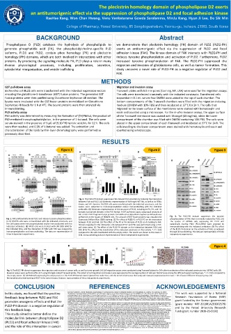
. Figure 1 Figure 2 Figure 3
A B C D
A B
A B
E F G H
C
D E
Fig. 2. The PLD2-PH domain suppresses FAK-induced PLD activation by reducing the interaction
between FAK and PLD2. (A) Schematic representation of full-length WT FAK, as well as its FERN,
kinase, and FAT domains. HEK293 cells were transfected with the indicated constructs, and the
lysates were subjected to immunoprecipitation and/or immunoblotting with the indicated
antibodies. (B) Schematic representation of the structure of PLD2, including its PX, PH, and I-IV
domains (conserved domains in the PLD family). A GST pull-down assay using equal amounts of
GST or GST-PLD2 fragment fusion proteins immobilized on Glutathione Sepharose 4B beads was
performed on the lysates of HEK293 cells. The amount of GST fusion proteins was visualized by Fig. 3. The PLD2-PH domain suppresses the tyrosine
Fig. 1. FAK preferentially binds PLD2 and induces tyrosine phosphorylation. Coomassie Brilliant Blue (CBB) staining. (C) The effect of PLD2-F1 or PLD2-F2 on the interaction phosphorylation of FAK that is normally induced by PLD2. (A)
(A-D) HEK293 cells were co-transfected with the indicated constructs, and between PLD2 and FAK. (D) The effect of the PX or PH domains of PLD2 on its binding to FAK. The lysates of U87MG cells expressing WT PLD2 and
the cell lysates were immunoprecipitated and/or immunoblotted with the Domain mapping for the interaction site between PLD2 and FAK was performed using a GST- catalytically inactive PLD2 (PLD2-KRM) were analyzed by
indicated antibodies. (E) HN33 cells were treated with LPA (100 mM) for pull down assay. (E) The effect of the PLD2-PH domain on the interaction between PLD2 and immunoblotting with the indicated antibodies. (B) The effect
the indicated time, and the interaction of PLD2 with FAK was analyzed by FAK. (F-H) The effect of the transfection of the indicated constructs on PLD activity. *, P < 0.05 of the PLD2-PH domain on the activation of FAK, as analyzed
immunoprecipitation and immunoblotting. The data are representative of compared to the cells transfected with the empty vector. The results are shown as the mean ± through immunoblotting. The data are representative of three
three independent experiments. SEM. Immunoblottingdata are representativeof three independent experiments independent experiments.
.
A B D Figure 4
C
Fig. 4. The PLD2-PH domain suppresses the migration and invasion of cancer cells, as well as tumor growth. (A) Cell migration assays were conducted using Transwell plates for 24 h after transfection of the indicated constructs into U87MG cells. (B)
Invasion assays were performed for 24 h using Matrigel-coated Transwell plates. The extent of cell migration and invasion was expressed as the average number of cells per field of view among the different experimental groups. *, P < 0.05 compared to
the empty vector. (C) U87MG cells expressing the empty vector or the PLD2-PH domain were injected into nude mice, and the tumor volume was then analyzed. Data are expressed as the mean ± SD of seven different mice per group. (D)
Immunohistochemicalstaining of Ki67 in the tumor tissues. Data are representative of three independent experiments.
CONCLUSION REFERENCES ACKNOWLEDGEMENTS
In this study, we found that the positive [1]Gomez-Cambronero, J. (2014). Phosphatidic acid, phospholipase D and tumorigenesis. Advances in biological regulation 54, 197-206.[2]Bruntz, R.C., Lindsley, C.W. and Brown, H.A. (2014). This work was supported by a National
Phospholipase D signaling pathways and phosphatidic acid as therapeutic targets in cancer. Pharmacological reviews 66, 1033-1079.[3]Nelson, R.K. and Frohman, M.A. (2015). Physiological and
feedback loop between PLD2 and FAK pathophysiological roles for phospholipase D. Journal of lipid research 56, 2229-2237.[4]Ahn, B.-H. and Jo, Y.-H. (2001). Differential tyrosine phosphorylation of phospholipase D isozymes by Research Foundation of Korea (NRF)
hydrogen peroxide and the epidermal growth factor in A431 epidermoid carcinoma cells. Molecules & Cells (Springer Science & Business Media BV) 11[5]Kang, D.W. and Choi, K.-Y. (2014). Functional
regulation of phospholipase D expression in cancer and inflammation. Journal of Biological Chemistry 289, 22575-22582.[6]Zhao, C., Du, G., Skowronek, K., Frohman, M.A. and Bar-Sagi, D. (2007).
promotes oncogenic effects and that the Phospholipase D2-generated phosphatidic acid couples EGFR stimulation to Ras activation by Sos. Nature cell biology 9, 707.[7]Frohman, M.A. (2015). The phospholipase D superfamily as therapeutic grant funded by the Korean government
targets. Trends in pharmacological sciences 36, 137-144.
[8]Slaaby, R., Jensen, T., Hansen, H.S., Frohman, M.A. and Seedorf, K. (1998). PLD2 complexes with the EGF receptor and undergoes tyrosine phosphorylation at a single site upon agonist stimulation.
PLD2-PH domain is a negative regulator of Journal of Biological Chemistry 273, 33722-33727.[9]Ahn, B.-H. et al. (2003). Transmodulation between phospholipase D and c-Src enhances cell proliferation. Molecular and cellular biology 23, 3103- (grant number NRF-2018R1A2B3002179)
3115.[10]Brown, H.A., Thomas, P.G. and Lindsley, C.W. (2017). Targeting phospholipase D in cancer, infection and neurodegenerative disorders. Nature reviews Drug discovery 16, 351.[11]Jang, J.-H.,
and by the Yonsei University Research
this feedback loop. Lee, C.S., Hwang, D. and Ryu, S.H. (2012). Understanding of the roles of phospholipase D and phosphatidic acid through their binding partners. Progress in lipid research 51, 71- Fund (grant number 2019-22-0193)
81.[12]Gomez-Cambronero, J. (2012). Biochemical and cellular implications of a dual lipase-GEF function of phospholipase D2 (PLD2). Journal of leukocyte biology 92, 461-467.[13]Park, M.H., Choi, K.-
Y., Jung, Y. and Min, D.S. (2014). Phospholipase D1 protein coordinates dynamic assembly of HIF-1α-PHD-VHL to regulate HIF-1α stability. Oncotarget 5, 11857.[14]Henkels, K.M., Peng, H.-J., Frondorf,
This study aimed to better define the K. and Gomez-Cambronero, J. (2010). A comprehensive model that explains the regulation of phospholipase D2 activity by phosphorylation-dephosphorylation. Molecular and cellular biology 30,
2251-2263.[15]Schaller, M.D. and Parsons, J.T. (1995). pp125FAK-dependent tyrosine phosphorylation of paxillin creates a high-affinity binding site for Crk. Molecular and cellular biology 15, 2635-
2645.[16]Park, M.H., Ahn, B.-H., Hong, Y.-K. and Min, D.S. (2009). Overexpression of phospholipase D enhances matrix metalloproteinase-2 expression and glioma cell invasion via protein kinase C
molecular link between phospholipase D2 and protein kinase A/NF-κB/Sp1-mediated signaling pathways. Carcinogenesis 30, 356-365.[17]Elvers, M. et al. (2010). Impaired αIIbβ3 integrin activation and shear-dependent thrombus formation
in mice lacking phospholipase D1. Sci. Signal. 3, ra1-ra1.[18]Min, D., Ahn, B.-H., Rhie, D.-J., Yoon, S.-H., Hahn, S., Kim, M.-S. and Jo, Y.-H. (2001). Expression and regulation of phospholipase D during
(PLD2) and focal adhesion kinase (FAK) neuronal differentiation of PC12 cells. Neuropharmacology 41, 384-391.[19]Bligh, E.G. and Dyer, W.J. (1959). A rapid method of total lipid extraction and purification. Canadian journal of
biochemistry and physiology 37, 911-917.[20]Schaller, M.D., Hildebrand, J.D., Shannon, J.D., Fox, J.W., Vines, R.R. and Parsons, J.T. (1994). Autophosphorylation of the focal adhesion kinase,
pp125FAK, directs SH2-dependent binding of pp60src. Molecular and cellular biology 14, 1680-1688.[21]Mitra, S.K., Hanson, D.A. and Schlaepfer, D.D. (2005). Focal adhesion kinase: in command and
and the role of this interaction in cancer. control of cell motility. Nature reviews Molecular cell biology 6, 56.[22]Lim, Y., Han, I., Jeon, J., Park, H., Bahk, Y.-Y. and Oh, E.-S. (2004). Phosphorylation of focal adhesion kinase at tyrosine 861 is
crucial for Ras transformation of fibroblasts. Journal of Biological Chemistry 279, 29060-29065.[23]Subauste, M.C., Pertz, O., Adamson, E.D., Turner, C.E., Junger, S. and Hahn, K.M. (2004). Vinculin
modulation of paxillin–FAK interactions regulates ERK to control survival and motility. The Journal of cell biology 165, 371-381.[24]Schwock, J., Dhani, N. and Hedley, D.W. (2010). Targeting focal
adhesion kinase signaling in tumor growth and metastasis. Expert opinion on therapeutic targets 14, 77-94.