
Page 35 - D. Cancer biology
P. 35
Inhibition of phospholipase D2 augments histone
deacetylase inhibitor-induced cell death in breast cancer
cells
Do Sik Min¹, MinJu Kang¹, Won Chan Hwang¹, Venu Venkatarame Gowda Saralamma¹, JuWon Kim¹,
RaeHee Kang¹, Hyun Ji Lee¹
¹College of pharmacy, Yonsei University, InCheon 21983, Republic of Korea
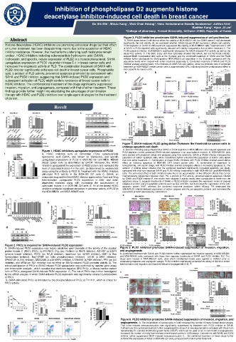
Abstract Figure 3. PLD2 inhibition accelerates SAHA-induced suppression of cell proliferation.
A, SAHA doses below 2 mM did not affect the viability of MDA-MB231 cell, but SAHA above 5 mM decreased
significantly the cell viability. B, we examined whether SAHA-induced PLD2 expression affects cell viability.
Histone deacetylase (HDAC) inhibitors are promising anticancer drugs but their effect PLD2 depletion or SAHA (5 mM) treatment suppressed the viability of MDA-MB231 cells.Treatment with 5 mM
on tumor treatment has been disappointing mainly due to the acquisition of HDAC of SAHA in PLD2-depleted cells significantly reduced cell viability compared to that of either treatment C, The
inhibitor resistance. However, the mechanisms underlying such resistance remain PLD2-selective inhibitor, VU0285655-1, significantly decreased the SAHA-induced PLD activity as well as the
basal PLD activity. D. The BrdU assay with flow cytometry showed that SAHA, but not the PLD2 inhibitor,
unclear. HDAC inhibitors including suberoylanilide hydroxamic acid (SAHA), reduced BrdU incorporation into cells at the S-phase and the combined treatment of SAHA and the PLD2
trichostatin, and apicidin, induce expression of PLD2 in a transcriptional level. SAHA inhibitor further decreased the BrdU-positive MDA-MB231 cell population in the S-phase compared with the
upregulates expression of PLD2 via protein kinase C-z in breast cancer cells and population levels when treated with either treatment separately E, Combined treatment of SAHA and PLD2
inhibitor showed approximately 10% inhibition on MCF-10A cell viability, while the inhibition rate of combined
increases the enzymatic activity of PLD. The combination treatment of SAHA with treatment on MDA-MB231 breast cancer cells is approximately 60%, indicating that the combinational effect is
PLD2 inhibitor significantly enhances cell death in breast cancer cells. Phosphatidic more specific for cancer cells
acid, a product of PLD activity, prevented apoptosis promoted by cotreatment with
SAHA and PLD2 inhibitor, suggesting that SAHA-induced PLD2 expression and
subsequent activation of PLD2 might confers resistance of breast cancer cells to
HDAC inhibitor. The combinational treatment of the drugs significantly suppressed
invasion, migration, and angiogenesis, compared with that of either treatment. These
findings provide further insight into elucidating the advantages of combination
therapy with HDAC and PLD2 inhibitors over single-agent strategies for the treatment
of cancer.
Result
Figure 4. SAHA-induced PLD2 upregulation increases the threshold for cancer cells to
undergo apoptotic cell death
A, Annexin V binding assay showed that SAHA or PLD2 depletion in MDA-MB-231 cells induced apoptosis and
Figure 1. HDAC inhibitors upregulate expression of PLD2 PLD2-depletion further promoted SAHA-induced apoptosis in an accumulative manner. B, MDA-MB-231 cells
A, HDAC inhibitors such as trichostatin (TSA), suberoylanilide were stained with propidium iodide and analyzed using flow cytometry. SAHA or PLD2 inhibitor increased the
hydroxamic acid (SAHA, also known as Vorinostat), and apicidin population of subG1 apoptotic cells, while cotreatment further enhanced the population of subG1 cells above
upregulated expressions of PLD2 in MDA-MB 231 and MDA- MB435 that from either treatment. C, Combination of these HDAC inhibitors with PLD2 inhibitor showed accumulative
breast cancer cells as determined by q-PCR. Moreover, the HDAC effect on inducing apoptosis of MDA-MB 2312 breast cancer cells D, combination of rapamycin or
inhibitors upregulated the expression of PLD2 protein and increased the temozolomide, anti-cancer drugs, with PLD2 inhibitor exerted synergistic effects in increasing apoptosis E, The
level of acetylated histone 4 in the cells, as determined by western blot annexin V binding assay showed that the combined treatment significantly increased apoptotic cell death,
assay using the antibody to PLD2 B, Treatment with the HDAC inhibitors compared with that from separate SAHA and PLD2 inhibitor treatments in both MDA-MB-231 and MDA-MB435
stimulated PLD activity in the MDA-MB 231 cells C, SAHA, an cells. The enhanced effect of both inhibitors looks like to be accumulation of two different effects that can be
anticancer drug and the first HDAC inhibitor approved by Food and Drug mechanistically independent between them. PA, a product of PLD activity, protected against apoptosis induced
by SAHA and PLD1 inhibitor F, the results from caspase 3 activity assay were comparable to those from the
Administration ,upregulated PLD2 expression in time- and dose- apoptosis assay G, separate treatments of SAHA or PLD2 inhibitor increased the expression levels of cleaved
dependent manners along with increasing the accumulation of caspase-3 protein and pro-apoptotic proteins such as Bax and Bim, but decreased the expression of the anti-
acetylated histone 4 in MDA-MB 231 cells D, All of the tested HDAC apoptotic protein XIAP, whereas the combined treatment produced further efficacy. PA decreased the
inhibitors produced significant increases in promoter activity of PLD2 in SAHA/PLD2 inhibitor-induced expression of active caspase and the pro-apoptotic proteins and recovered the
the MDA-MB231 and MDA-MB435 cells expression of XIAP decreased by cotreatment
Figure 2. PKCz is required for SAHA-induced PLD2 expression
A, SAHA-induced PLD2 expression was largely abolished upon blockade of the activity of the atypical Figure 5. PLD2 inhibition promotes SAHA-induced suppression of invasion, migration,
protein kinase C (PKC), PKCz, by PS-PKCz (Fig. 2a). Rottlerin (a PKCδ inhibitor), AG1487 (a EGFR and angiogenesis
tyrosine kinase inhibitor), PDTC (an NF K B inhibitor), rapamycin (an mTOR inhibitor), B581 (a Ras A, Combined treatment with SAHA and PLD2 inhibitor further suppressed the invasive capacity of MDA-MB-231
farnesylation inhibitor), Bay117085 (an IκBα phosphorylation inhibitor), U0126 (a MEK inhibitor), and MDA-MB435 cells, compared with those from separate treatments of SAHA and PLD2 inhibitor. B,C The
SP600125 (a JNK inhibitor), SB203580 (a p38 MAPK inhibitor), LY294002 (a PI3K inhibitor), PP2 (an Src drugs were treated in MDA-MB-231 cells, after which conditioned media were applied to HUVEC prior to
inhibitor), and MTM (an Sp1 inhibitor) had no effect on SAHA-induced PLD2 promoter activity. B, The undertaking migration and angiogenic assays. PLD2 inhibition significantly enhanced the ability of SAHA to inhibit
tube formation and migration, an important feature of angiogenesis D, E,
critical participation of PKCz in SAHA-induced PLD2 upregulation was confirmed by reporter gene assay
and immunoblot analysis , which revealed that dominant-negative (DN) PKCz, a kinase-inactive mutant
form of PKCz, abrogated SAHA-induced PLD2 expression. C, The role of PKCz was further investigated
by the siRNA analysis in which SAHA-induced PLD2 expression was significantly reduced by knockdown
of PKCz
D, SAHA stimulated PKCz as indicated by the phosphorylation of PKCz at Thr 410 , which is critical for
PKCz activity
Figure 6. PLD2 inhibition promotes SAHA-induced suppression of invasion, migration, and
angiogenesis. A, The implantation of cancer cells in CAM increased the number of newly formed blood vessels.
That tumor-induced neovascularization was significantly suppressed by treatment with PLD2 inhibitor or SAHA.
Furthermore, the combined treatment further suppressed the amount of neovascularization compared with those from
monotherapy B, vascular endothelial growth factor (VEGF), which can be used as an in vivo model of angiogenesis,
increased the number of newly formed blood vessel branch points in CAM assays. Cotreatment with SAHA and PLD2
inhibitor further suppressed VEGF-induced neovascularization C, , the combined treatment of these drugs further
inhibited the expression of VEGF in MDA-MB-231 cells, compared with that of either treatment