
Page 167 - D. Cancer biology
P. 167
Hepatitis B virus X protein enhances liver cancer cell migration by regulating
calmodulin-associated actin polymerization
Mi-jee Kim, Inho Kang, and Jeong Keun Ahn*
Department of Microbiology & Molecular Biology, Chungnam National University, Daejeon, Korea
BACKGROUND AIM
Hepatitis B virus (HBV) is one of the major pathogens of acute hepatitis, chronic hepatitis, cirrhosis, and hepatocellular carcinoma (HCC). Recently, it has been estimated that about 53% of HCC cases To understand the role of HBx in the development
in the world are related to HBV. There are many possible mechanisms whereby HBV may cause HCC. However, the exact mechanism by which HBV infection leads to liver cancer remains unclear. of HCC, we attempted to find cellular proteins
HBV X protein (HBx) is a small regulatory protein is required for the establishment of viral replication the development of HCC. HBx also promotes cellular invasiveness, adhesion, migration, and interacting with HBx by yeast two-hybrid screening
morphogenesis by regulating adhesion proteins, matrix metalloproteinases, and cytoskeletal proteins. Suggesting that HBx protein is associated with HCC metastasis. Calmodulin (CaM) is a highly analysis. Interestingly, CaM was identified to bind
conserved calcium binding protein that contains four EF-hand calcium binding motif. CaM acts as a major calcium sensor and relays the calcium signaling. Recently, it was reported that CaM is HBx. Here, we report that the HBx regulates CaM
associated with cancer formation such as angiogenesis, metastasis, and cell migration. Cell migration is an important cellular process for cancer metastasis. F-actin polymerization and actin and subsequently affects actin cytoskeleton
cytoskeletal rearrangement play pivotal roles in the migration of cells. Cofilin is a major actin depolymerizing factor which is regulated by LIMK1-mediated phosphorylation. Inacivated reorganization and cell migration associated with
phosphorylated cofilin (p-cofilin) induces actin polymerization and enhances cell migration. The stability of LIMK1 is regulated by HSP90 that promotes LIMK1 homo-dimerization and trans- HCC metastasis.
phosphorylation.
METHODS
Cells culture Human hepatocellular carcinoma cell lines including HepG2, and HepG2.2.15, Chang human liver cells and HEK 293T cells were maintained in DMEM supplemented with 10% heat-inactivated FBS and 1% penicillin-streptomycin-
amphotericin B mixture at 37℃ in humidified atmosphere with 5% CO2. Plasmids and antibodies The Flag-HBx wes a kind gift of Dr. S. Kim. The vector encoding the HBx-siRNA (adr) was a kind gift from Dr. Oi-lin Ng. The vectors encoding Flag-
HSP90 and myc-LIMK were gifts of Dr. J. Kim. GFP-LIMK and GFP-LIMK mutant plasmids ware a kind gifts of Dr. Bernard. Calmodulin 3 full gene was cloned into the mammalian expression vector, PEBG to express GST–CaM. The point-mutant of
HBx protein was generated by changing amino acid from lysine to alanine in the calmodulin binding motif of HBx protein. The antibodies were used as followings: anti-HBx, anti-HSP90, anti-LIMK, anti-cofilin, anti-GFP, anti-Myc, anti-α-tubulin, anti-p-
cofilin, anti-GST, anti-Flag. Immunoprecipitation and GST pull down assay Cells were transfected with plasmids and incubates for 48hr. Cells were lysed with modified RIPA buffer for 30min at 4℃. The cleared lysate wes incubated with the indicated
antibody for overnight (o/n) at 4℃, and then incubated with protein A-conjugated beads for 2 hr at 4℃. The beads were washed several times with modified RIPA buffer. The pellets were added to SDS sample buffer and boiled for 10min. The samples were
resolved on SDS-PAGE for immunoblotting. GST-pull down assay was carried out using glutathione-Sepharose 4B beads. Western blot analysis Cell lysates were resolved on SDS-PAGE and the proteins in the gels were transferred onto a PVDF
membrane. The membranes were incubated with 5% (w/v) skim milk in PBSt (PBS containing 0.2% Tween 20) and then reacted with primary antibodies. After washing three times with PBSt, the membranes were incubated with horseradish peroxidase-
conjugated anti-IgG. After washing three times with PBSt, the proteins were detected with the ECL reagent (Millipore). Immunofluorescence microscopy Cells were splited on sterile galss cover silps and incubated for o/n. 48hr after transfection, cells
were washed with cold PBS and fixed for 10 min with 4% formaldehyde. Cells were washed three times with cold PBS and permeabilized with PBSt for 5min. Cells were incubated with primary antibodies in PBSt (with 0.2% BSA) for o/n and followed by
incubation for 1h with fluorochrome-conjugated secondary antibodies. The fluorescence was examined using a fluorescence microscope. Wound healing assay After 24h transfection, cells were grown to 80% confluence in complete DMEM. Monolayers
were wounded by scratching with a sterile plastic 20 µL micropipette tip, washed with PBS, and incubated in DMEM with 0.1% FBS. After 24 h, cells were fixed with 4% paraformaldehyde in PBS for 5 min at room temperature and photographed using a
microscope. The extent of migration into the wound area was evaluated quantitatively using Image J software. Cell migration assay Migration assays were carried out with a QCM chemotaxis cell migration assay kit (Chemicon). Cells were added to the
upper chambers and the chambers were placed in medium containing dish. Migration assays were carried out for 24hr. Migratory cells on the bottom of the insert membrane are dissociated from the membrane using Cell Detachment buffer. Dissociated cells
were attached to by CyQuant GR dye. The green fluorescences were detected with fluorometer at 480/520 nm. Tail vein injectioln A suspension of 100μl buffer solution containing 1×10 6 B16F10 cells was injected into the tail vein of 6-week-old female
nude mouse (Balb/c-nu). The animals were sacrificed at 21 days after injection of melanomal cell.
RESULTS
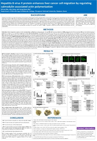
HBV X protein binds to calmodulin To identify the cellular proteins interacting with
HBx, we performed a yeast two hybrid screening. As a result, 14 cDNA clones had
positive interaction with HBx. One of them was calcium modulate protein (CaM). The
interaction of HBX and CaM was further demonstrated by a GST pull down assay in
HEK 293T cells (Fig.1a) and HepG2 cells (Fig.1b). We also tested the calcium
dependency of the HBx-CaM binding by using CaCl 2 or EGTA (Fig.1c). It turned out
that HBx interacts with CaM in a calcium dependent manner. In addition, we generated
the point-mutant plasmid of HBx to changing one amino acid as in putative CaM
binding motif of HBx protein. Interestingly, we found that HBx mutant can’t bind with
CaM (Fig.1d).
HBV X protein increases cell migration and F-actin polymerization. Recently, it
has been shown that CaM regulates the several small GTPases. Small GTPases play
essential role in cytoskeleton remodeling associated with cell motility. We tested the
effects of HBx and CaM on actin cytoskeleton reorganization in hepatocytes as
assessed by immuno-fluorecence microscopy (Fig.2a) and wound healing assays
(Fig.2b). The actin polymerization leading to stress fiber formation was reduced by
CaM and elevated by HBx. The wound healing assays also indicates that CaM
decreases cell migration, while HBx enhances cell migration conversely. In addition,
liver cells expressing HBx exhibited a much greater ability to migrate that the cells
expressing HBx mutant devoid of CaM binding motif. (Fig.2c).
HBV X protein increases cofilin phosphorylation Cofilin is a ubiquitous actin-
binding factor required for the reorganization of actin filaments and is inactivated by
phosphorylation reaction of LIM-kinase. To determine whether HBx has an effect to
modulate p-cofilin, pSuper-shHBx plasmid was transfected into HepG2.215 cells
which produced HBV virus particles (Fig.3a). Interestingly, the level of p-cofilin was
decreased when HBx expressing abrogated. In previous study, LIMK1 has been
shown to affect cell motility, invasion, and cancer metastasis. The activity and the
stability of LIMKs are further regulated by HSP90 that promotes LIMK1 homo-
dimerization and trans-phosphorylation. Therefore, we investigated whether HSP90
and LIMK are involved in HBx-mediated cofilin phosphorylation. Actually, both HBx
and CaM only regulate p-cofilin level without changing the levels of HSP90 an LIMK
(Fig.3b,c). In addiation, we found that HBx rescued p-cofilin level which was
repressed by CaM without affecting HSP90 and LIMK (Fig.3d,e,f).
HBV X protein interferes the binding between CaM and HSP90. Previous studies
showed that Hsp90 binds to CaM in a Ca 2+ -dependent manner, and HSP90 is a
constitutive dimer where each monomer consists of three domains. The CaM-binding
site in Hsp90 is a short peptide in the C-terminal part of the protein, which was also
required for HSP90 dimerization. For these reasons, we speculated that the binding of
HBx to CaM might regulate the binding between CaM and HSP90. As predicted, the
dimerization of HSP90 was significantly reduced by CaM (Fig.4a), while HBx
interferes with the binding between CaM and HSP90 (Fig.4b). Since, HSP90
associates with LIMK1 and increases the activity and the stability of LIMK, we tested
the effects of CaM and HBx on the interaction between HSP90 and LIMK by
immunoprecipitation assay in HEK293T cells (Fig.4c) and HepG2 cells (Fig.4d). As a
result, the interaction between HSP90 and LIMK was repressed by CaM, however the
reduced binding was significantly rescued by HBx. These data indicate that the
interaction between HBx and CaM regulates the p-cofilin level through HSP90-
LIMK1 pathway. HBx interferes the binding between CaM and HSP90, and elevate
HSP90-LIMK dimerization to activate LIMK.
HBV X protein increases tumor metastasis in vivo. To further explore the
metastatic potential of HBx-CaM interaction in vivo, we performed intravenously
injecting cancer cells expressing either HBx or HBx mutant into nude mice. A
suspension of B16F10 cells ware injected into the tail vein of 6-week-old female nude
mouse (Fig.5); the first group was injected with B16F10 cells (a), the second group
was injected with HBx expressing B16F10 cells (b), the third group was injected with
HBx mutant expressing B16F10 cells (c). Interestingly, we observed that HBx induced
lung metastasis significantly, while HBx mutant exhibited less metastatic portion.
CONCLUSION REFERENCES
- HBV X protein binds to calmodulin in a calcium ion-dependent manner. 1. Lupberger J, Hildt E. (2007) "Hepatitis B virus-induced oncogenesis." World J Gastroenterol
2. Wang XW, Hussain SP, Huo TI, Wu CG, Forgues M, Hofseth LJ, Brechot C, Harris CC. (2002) "Molecular pathogenesis of human hepatocellular carcinoma."
- HBV X protein increases cell migration and F-actin polymerization. Toxicology
3. Zhang X, Zhang H, Ye L. (2006) "Effects of hepatitis B virus X protein on the development of liver cancer." J Lab Clin Med
- HBV X protein increases cofilin phosphorylation.
4. Kedrin D, van Rheenen J, Hernandez L, Condeelis J, Segall JE. (2007) "Cell motility and cytoskeletal regulation in invasion and metastasis." J Mammary Gland Biol
Neoplasia.
- HBV X protein interferes the binding between CaM and HSP90. Also,
HBV X protein increases the binding between HSP90 and LIMK1, but 5. Alessandro R, Masiero L, Liotta LA, Kohn EC. (1996) "The role of calcium in the regulation of invasion and angiogenesis." In Vivo Contact information
CaM decreases the binding between HSP90 and LIMK1. 6. Buchholz M, Ellenrieder V. "An emerging role for Ca2+/calcineurin/NFAT signaling in cancerogenesis." (2007) Cell Cycle
7. Wang W, Condeelis JS. (2006) "The activity status of cofilin is directly related to invasion, intravasation, and metastasis of mammary tumors." J Cell Biol
- HBV X protein increases tumor metastasis in vivo.
8. Wang W, Eddy R, Condeelis J. (2007) "The cofilin pathway in breast cancer invasion and metastasis." Nat Rev Cancer Address correspondence to: Jeong Keun Ahn,
HBV X protein regulates F-actin rearrangement associated with cell 9. Ora Bernard, (2007) "Lim kinases, regulators of actin dynamics", Biochemistry & Cell Biology Dept. of Microbiology & Molecular Biology,
migration of HCC through its interaction with CaM. In this study, we 10. Michiru Nishita, (2005) "Spatial and temporal regulation of cofilin activity by LIM kinase and Slingshot is critical for directional cell migration.", The Journal of Cell School of Bioscience and Biotechnology,
elucidate the regulatory mechanism of cell migration induced by HBx Biology Chungnam National University, Daejeon, Korea.
which controls CaM acting as a regulatory protein of liver cancer 11. Ora Bernard, (2006) “Hsp90 increases LIM kinase activity by promoting its homo-dimerization”, The FASEB Journal
metastasis. 12. Daniel N. Bolon, (2007) “Dimerization of Hsp90 Is Required for in Vivo Function”, THE JOURNAL OF BIOLOGICAL CHEMISTRY E-mail: jkahn@cnu.ac.kr