
Page 127 - D. Cancer biology
P. 127
Effects of pregnenolone on the human breast cancer cells
1
1
1
1
1
1
Min Jae Kim , Sung-Min An , So Young Kim , Da Som Kim , Da Hee Kang , and Beum-Soo An *
1 Department of Biomaterials Science, College of Natural Resources & Life Science/Life and Industry Convergence Research Institute,
Pusan National University, Miryang, Gyeongnam, Republic of Korea
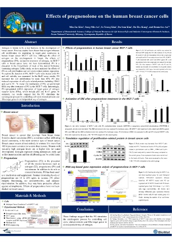
Abstract Results
Estrogen is known to be a key hormone in the development of Effects of pregnenolone in human breast cancer MCF-7 cells
breast cancer. Previous studies have shown that estrogen enhances A Figure 1. (A) Cell proliferation and viability was analyzed by
cell proliferation via regulating its target gene expression in BrdU and MTT assays after 24 h after treatment of Con, E2 and
human breast cancer MCF-7 cell line. Although the effect of PG on MCF-7 cell. (B) Transcription levels of ER α target gene
estrogen on the carcinogenesis in breast, the effects of such as pS2 and Ki-67 were analyzed by real-time PCR. (C) MCF-
pregnenolone (PG), an inactive precursor of estrogen, on MCF-7 7 cells transfected with control and siRNA against ER α and
cells or breast cancer have not been demonstrated. PG is a transcription levels of ER α target gene were analyzed by real-time
PCR. (D) After treatment of MCF-7 cells with E2 and PG,
precursor in the biosynthesis of most of the steroid hormones medium was harvested. E2 concentration in cell growth medium
containing estrogen. In this study, we have analyzed the effects of was measured by ELSIA assay. Data were expressed as the mean
PG on cell proliferation and cell cycle-related protein expression. ± SD. * P<0.05, *** P<0.005 compared to the control group.
To explore the function of PG, MCF-7 cells were treated with PG
and cell viability was examined. In the BrdU assay results, PG B C D
increased the cell proliferation by more than 40%. PG also
induced expression of cell cycle related-proteins including CDK2
and cyclin D1. To reveal the mechanism of PG, we performed
RNA seq after treatment of PG in the MCF-7 cells. Interestingly,
PG upregulated mRNA expression of target genes of estrogen
receptor alpha (ERα), which include pS2 and Ki-67 genes. In
summary, our results suggest that the PG stimulates the
carcinogenic process by controlling cell cycle related-proteins and
ERα target genes in an independent way of estrogen. Activation of ERE after pregnenolone treatment in the MCF-7 cells
A B C
Introduction
Breast cancer
Among them, drug-
loaded dissolving MNs
are made of dissolving Figure 1. (A) After treatment of MCF-7 cells with PG, methylpiperidino pyrazole (MPP/ER α antagonist), pyrazolo[1,5-α]pyrimidines (PHTPP/ER β
antagonist), protein were harvested. The ERE activation levels were analyzed by luciferase assay. (B) MCF-7 cells transfected with control and siRNA against
ER α and ERβ and the ERE activation levels were analyzed by luciferase assay. (C) Activation of ERE was measured on E2 and PG-cotreated MCF-7 cells.
materials suchcalled infiltrating Translation regulation of proliferation-related protein in breast cancer cells
as
Breast cancer is cancer that develops from breast tissue.
Data were expressed as the mean ± SD. * P<0.05 compared to the control group.
Invasive ductal carcinoma (IDC), sometimes
ductal carcinoma, is the most common type of breast cancer.
carboxymethyl cellulose, A B Figure 3. Total protein was harvested from MCF-7 cells
Breast cancer occurs almost entirely in women. It is more than
100 times more common in women than in men. Women with
treated with PG. Translational levels of ER α (A) and gene
hyaluronic acid (HA), related to proliferation such as CDK , CDK4 and Cyclin
naturally high estrogen levels are at higher risk for tumor
development. Estrogen exposure during menopause, early age
D1 (B) were analyzed by western blot assay compared to
at first menstrucation and late childbearing can be a risk factor.
and chitosan, and the control group. Gene expression levels were normalized
Pregenolone
to the levels of β-actin. Data were expressed as the mean
Pregnenolone (PG) is the precursor
prepared of all the steroid hormones such as ± SD. * P<0.05 compared to the control group.
estrogen (E2), progesterone (P4) and
by RNA seq-based gene expression anlysis of pregnenolone in MCF-7 cells
testosterone. In addition to its role as
micromolding or A B PG_vs_Con Figure 3. (A) Heatmap showing the MCF-7 up-
a natural hormone, PG has been used
as a medication and supplement. Normal circulating levels of
and down-regulated genes for each biological
dwelling process with PG replicate. Differential expression analyses
pregnenolone are 10 to 230 ng/dL in women. PG affect
synaptic functioning, are neuroprotective, and enhance
identified 147 MCF-7 treated with PG up-
myelinization. In addition, they may have protective effects
drug/polymer solution. regulated and 93 MCF-7 treated with PG down-
against schizophrenia. Effects of pregnenolone have not been
regulated genes (log2 fold change > 1, p < 0.01)
studied on breast cancer.
showing differential gene expression between
C Con with high reproducibility. (B) Scatter plot
Materials & Methods MCF-7 treated with PG and control gruop. (C)
PG/Con Graph showing the number of up- and down-
regulated genes.
Cells
Michigan Cancer Foundation-7 cell (MCF-7 cell)
Experimental Methods
In vitro (MCF-7 cell) Conclusion Reference
Bromodeoxyuridine assay
MTT assay These findings suggest that the PG stimulates [1] Lottering, Mona-Liza, Marianne Haag, and Johanna C. Seegers. "Effects of 17β-estradiol metabolites on cell
cycle events in MCF-7 cells." Cancer Research 52.21 (1992): 5926-5932.
Transfection using siRNA of estrogen the carcinogenic process by controlling cell [2] Yamaga, Ryonosuke, et al. "RNA sequencing of MCF-7 breast cancer cells identifies novel estrogen-
receptor and ERE luciferase vector responsive genes with functional estrogen receptor-binding sites in the vicinity of their transcription start sites."
Western blot cycle related-proteins and ERα target genes in Hormones and Cancer 4.4 (2013): 222-232.
RNA sequencing an independent way of estrogen. [3] Soto, Ana M., and Carlos Sonnenschein. "The role of estrogens on the proliferation of human breast tumor
cells (MCF-7)." Journal of steroid biochemistry 23.1 (1985): 87-94.