
Page 123 - D. Cancer biology
P. 123
Mesothelial-to-mesenchymal transition induced by macrophages plays a role in
ovarian cancer cell invasion
Ju-Yeon Choi , Jin-Hyeong Lee a, b , and Jung-Hye Choi a, b *
a
a Department of Life and Nanopharmaceutical Sciences, Kyung Hee University, Seoul, South Korea
b College of Pharmacy, Kyung Hee University, Seoul, South Korea
Abstract Results
Conclusion
Peritoneal dissemination is a common characteristic of ovarian
cancer metastasis. Recently, an increasing number of studies A B A
have suggested that peritoneal mesothelial cells turn into
carcinoma-associated fibroblasts through mesothelial-to- MeT5A M-MeT5A 5 MeT5A_vector
MeT5A_nc886
mesenchymal transition (MMT), which plays a role in ovarian 10 8 * 4 * *
cancer metastasis. We found that macrophages induce the MMT of 6 Relative gene expression normalized by β-actin 3 * * *
MeT5A mesothelial cells and the macrophage-stimulated MeT5A Cell invasion (relative to MeT5A) 4 2 * *
cells (M-MeT5A) became markedly invasive. In addition, TGF-β1 2 1
secreted from macrophages induces MMT, stimulates the invasion 0 MeT5A M-MeT5A 0 VEGF Neuropilin TGF-β Vimentin MMP2 Fibronectin α-sma
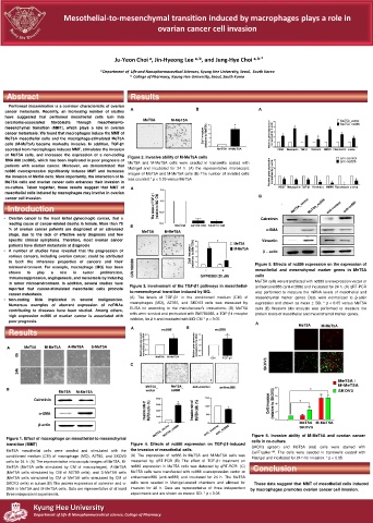
of MeT5A cells, and increases the expression of a non-coding Figure 2. Invasive ability of M-MeT5A cells
RNA 886 (nc886), which has been implicated in poor prognosis of MeT5A and M-MeT5A cells were seeded in transwells coated with 3 anti-control
anti-nc886
patients with ovarian cancer. Moreover, we demonstrated that Matrigel and incubated for 24 h. (A) the representative microscopic
nc886 overexpression significantly induces MMT and increases images of MeT5A and M-MeT5A cells (B) The number of invaded cells Relative gene expression 0 normalized by β-actin 2
the invasion of Met5A cells. More importantly, the interaction of M- was counted.* p < 0.05 versus MeT5A 1 * * * *
MeT5A cells and ovarian cancer cells enhances their invasion in * * *
co-culture. Taken together, these results suggest that MMT of A 1.5 VEGF Neuropilin TGF-β Vimentin MMP2 Fibronectin α-sma
mesothelial cells induced by macrophages may involve in ovarian
cancer cell invasion. 1.0 B
Introduction The levels of TGF-β1 (relative to MQ CM)
Introduction
• Ovarian cancer is the most lethal gynecologic cancer, that a 0.5 Calretinin
leading cause of cancer-related deaths in female. More than 75 0.0 MQ CM A2780 CM SKOV3 CM
% of ovarian cancer patients are diagnosed at an advanced B MeT5A M-MeT5A α-SMA
stage, due to the lack of effective early diagnosis and few
specific clinical symptoms. Therefore, most ovarian cancer 10 * Vimentin
patients have distant metastasis at diagnosis 8 MeT5A
• A number of studies have revealed that the progression of 6 M-MeT5A β – actin
various cancers, including ovarian cancer, could be attributed Cell invasion (relative to MeT5A) 4
to both the inherence properties of cancers and their 2 Figure 5. Effects of nc886 expression on the expression of
microenvironment. For example, macrophage (MQ) has been GW788388 0 - + mesothelial and mesenchymal marker genes in MeT5A
shown to play a role in tumor proliferation,
immunosuppression, angiogenesis, and metastasis by inducing GW788388 (20 µM) cells
in tumor microenvironment. In addition, several studies have MeT5A cells were transfected with nc886 overexpression vector or
reported that cancer-stimulated mesothelial cells promote Figure 3. Involvement of the TGF-β1 pathways in mesothelial- antisensenc886 (anti-nc886) and incubated for 24 h. (A) qRT-PCR
cancer metastasis. to-mesenchymal transition induced by MQ. was performed to measure the mRNA levels of mesothelial and
• Non-coding RNA implicated in several malignancies. (A) The levels of TGF-β1 in the conditioned medium (CM) of mesenchymal marker genes Data were normalized to β-actin
Numerous examples of aberrant expression of ncRNAs macrophages (MQ), A2780, and SKOV3 cells was measured by expression and shown as mean ± SD. * p < 0.05 versus MeT5A
contributing to diseases have been studied. Among others, ELISA kit according to the manufacturer's instructions. (B) MeT5A cells (B) Western blot analysis was performed to measure the
high expression nc886 of ovarian cancer is associated with cells were seeded and pretreated with GW788388, a TGF-β1 receptor protein levels of mesothelial and mesenchymal marker genes.
poor prognosis. inhibitor, for 2 h and incubated with MQ-CM * p < 0.05 A MeT5A M-MeT5A
Results A 2.0 nc886 * B 2.0 nc886 *
A MeT5A M-MeT5A A-MeT5A S-MeT5A Relative gene expression normalized by 18s rRNA 1.5 1.0 0.5 Relative gene expression normalized to 18s rRNA 1.5 1.0 0.5
0h 0.0 MeT5A M-MeT5A 0.0 Ctrl TGF-β1
C SKOV3
24h
MeT5A /
MeT5A_ MeT5A_ anti-control anti-nc886 8 M-MeT5A
B MeT5A M-MeT5A vector nc886 * SKOV3
Calretinin 800 * 150 Cell invasion (relative to SKOV3) 6 4 *
Invasion rate of MeT5A cells (%) 400 Invasion rate of MeT5A cells (%) 100 *
600
α-SMA 200 50 2
β-actin 0 0 0 MeT5A M-MeT5A
MeT5A_vector anti_control anti_nc886
MeT5A_nc886
SKOV3
Figure 1. Effect of macrophage on mesothelial-to-mesenchymal Figure 6. Invasive ability of M-MeT5A and ovarian cancer
cells in co-culture.
transition (MMT) Figure 4. Effects of nc886 expression on TGF-β1-induced SKOV3 (green) and MeT5A (red) cells were stained with
MeT5A mesothelial cells were seeded and stimulated with the the invasion of mesothelial cells. CellTracker TM . The cells were seeded in transwells coated with
conditioned medium (CM) of macrophage (MQ), A2780, and SKOV3 (A) The expression of nc886 in MeT5A and M-MeT5A cells was Matrigel and incubated for 24 h for invasion. * p < 0.05
cells for 24 h. (A) The representative microscopic images of MeT5A, M- measured by qRT-PCR (B) The effect of TGF-β1 treatment on
MeT5A (MeT5A cells stimulated by CM of macrophages), A-MeT5A nc886 expression in MeT5A cells was detected by qRT-RCR. (C) Conclusion
(MeT5A cells stimulated by CM of A2780 cells), and S-MeT5A cells MeT5A cells were transfected with nc886 overexpression vector or
(MeT5A cells stimulated by CM of MeT5A cells stimulated by CM of antisensenc886 (anti-nc886) and incubated for 24 h. The MeT5A
SKOV3 cells) in culture.(B) The protein expression of calretinin and α- cells were seeded in Matrigel-coated chambers and allowed for These data suggest that MMT of mesothelial cells induced
SMA in MeT5A and M-MeT5A cells. Data are representative of at least invasion for 48 h. Data are representative of three independent by macrophages promotes ovarian cancer cell invasion.
three independent experiments. experiments and are shown as mean± SD. * p < 0.05
Kyung Hee University
Department of Life & Nanopharmaceutical science, College of Pharmacy