


Page 320 - ebook
P. 320
1. Introduction                                         3. Results
       Implantable medical devices are used for support of regenerating and replacing  Characterization of microsphere
       organs and tissues in an incapable state. However, many people who use medical
       devices suffer various complications, such as medical device-related infections. The
       current approaches to prevent and treat these infections include disinfection of
       medical  devices  before  their  implantation  and  multiple-dose  systemic
       administration of antibiotics and. However, these solutions often result in a low
       effectiveness, short-term effect and chance of bacterial resistance. Importantly,
       bacteria can form biofilms on medical devices, which is effectively resistant to
       antibiotics. In this study, we aimed to devise a method of bio-inspiredly modifying
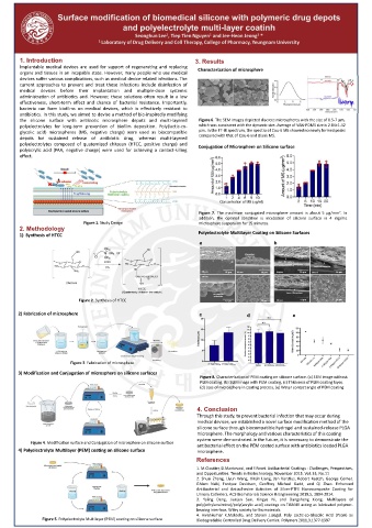
       the silicone surface with antibiotic microsphere depots and multi-layered  Figure 6. The SEM images depicted discrete microspheres with the size of 0.5-7 μm,
       polyelectrolytes for long-term prevention of biofilm deposition. Poly(lactic-co-  which was consistent with the dynamic size. Average of VAN-PDMS size is 2.84±1.42
       glycolic acid) microspheres (MS, negative charge) were used as biocompatible      . In the FT-IR spectrum, the spectra of Cou-6 MS showed no newly formed peaks
       depots for sustained release of antibiotic drug, whereas multi-layered  compared with that of Cou-6 and Blank MS.
       polyelectrolytes composed of quaternized chitosan (HTCC, positive charge) and  Conjugation of Microsphere on Silicone surface
       polyacrylic acid (PAA, negative charge) were used for achieving a contact-killing
       effect.


                                                                                                              2
                                                                 Figure 7. The maximum conjugated microsphere amount is about 5 μg/mm . In
                                                                 addition, the optimal condition is incubation of silicone surface in 4 mg/mL
                            Figure 1. Study Design               microsphere suspension for 15 minutes.
       2. Methodology
       1) Synthesis of HTCC                                      Polyelectrolyte Multilayer Coating on Silicone Surfaces
                                                                 a                       b


                                  (GTMAC)
                                   HClO 4


                          Figure 2. Synthesis of HTCC
                                                                               12 µm                 60µm
       2) Fabrication of microsphere                             c              d              e


                                                                                                    15 µm
                                                                               12 µm


                        Figure 3. Fabrication of microsphere
       3) Modification and Conjugation of microsphere on silicone surfaces
                                                                 Figure 8. Characterization of PEM coating on silicone surface. (a) SEM image without
                                                                 PEM coating, (b) SEM image with PEM coating, (c) Thickness of PEM coating layer,
                                                                 (d) Loss of microsphere in coating process, (e) Water contact angle of PEM coating


                                                                4. Conclusion
                                                                Through this study, to prevent bacterial infection that may occur during
                                                                medical devices, we established a novel surface modification method of the
                                                                silicone surface through biocompatible hydrogel and sustained-release PLGA
                                                                microsphere. The morphology and various characteristics of this coating
                                                                system were demonstrated.In the future, it is necessary to demonstrate the
           Figure 4. Modification surface and Conjugation of microsphere on silicone surface  antibacterial effect on the PEM coated surface with antibiotics loaded PLGA
       4) Polyelectrolyte Multilayer (PEM) coating on silicone surface  microsphere.
                                                                References
                                                                1. M.Cloutier, D.Mantovani, and F.Rosei. Antibacterial Coatings : Challenges, Perspectives,
                                                                and Opportunities. Trends in Biotechnology, November 2015. Vol. 33, No.11
                                                                2. Shuai Zhang, Liyun Wang, Xinjin Liang, Jan Vorstius, Robert Keatch, George Corner,
                                                                Ghlam Nabi, Fordyce Davidson, Geoffrey Michael Gadd, and Qi Zhao. Enhanced
                                                                Antibacterial and Antiadhesive Activities of Silver-PTFE Nanocomposite Coating for
                                                                Urinary Catheters. ACS Biomaterials Science & Engineering 2019,5, 2804-2814.
                                                                3. Yaling Deng, Jianjun Sun, Xingya Ni, and Dangsheng Xiong. Multilayers of
                                                                poly(ethyleneimine)/poly(acrylic acid) coatings on Ti6Al4V acting as lubricated polymer-
                                                                bearing interface. Wiley society for biomaterials
                                                                4. Hirenkumar K.Makadia, and Steven J.siegel. Poly Lactic-co-Glycolic Acid (PLGA) as
               Figure 5. Polyelectrolyte Multilayer (PEM) coating on silicone surface  Biodegradable Controlled Drug Delivery Carrier. Polymers 2011,3,1377-1397

