


Page 27 - ebook
P. 27
The potential anti-amyloidogenic candidate, SPA1413, for Alzheimer’s disease
60~70kDa
Young Kyo Kim , Shinwoo Kang , Hyewon Cho , Raok Jeon , Keun-A Chang 1,2,4*
1
3
3
2
1. Department of Health Sciences and Technology, Gachon Advanced Institute for Health Sciences & Technology, Gachon University, Incheon 21999, Korea
2. Neuroscience Research Institute, Gachon University, Incheon 21565, Korea
3. Research Institute of Pharmaceutical Sciences, College of Pharmacy, Sookmyung Women's University, Seoul, Republic of Korea
4. Department of Pharmacology, College of Medicine, Gachon University, Incheon 21999, Korea
• Correspondence: keuna705@gachon.ac.kr; Tel.: +82-32-899-6411
British Journal of Pharmacology, DOI: 10.1111/bph.15691, 2021 (IF: 8.739)
Background & Purpose Methods
Alzheimer’s disease (AD), the most common cause of neurodegenerative disorder, is characterized by the • Animals : 5xFAD (B6SJL) • Behavior test
deposition of amyloid-beta (Aβ) and neurofibrillary tangles. Recently, isoflavone derivatives have been shown Species: Mouse Novel object recognition test Y-maze test
to have neuroprotective effects against neurological disorders. For instance, genistein attenuated the Genes: APP, PSEN1 Habituation Adaptation Task
neuroinflammation and amyloid-β accumulation in Alzheimer's disease animal models, suggesting the Mutations: APP KM670/671NL (Swedish), APP I716V (Florida), APP V717I
(London), PSEN1 M146L (A>C), PSEN1 L286V
potential for use to prevent and treat Alzheimer's disease. In this study, the fifty compounds including Modification: APP: Transgenic; PSEN1: Transgenic
isoflavone derivatives were constructed and evaluated for their inhibitory effects on Aβ oligomerization and Disease Relevance: Alzheimer's Disease
Strain Name:B6SJL-Tg(APPSwFlLon,PSEN1*M146L*L286V)6799Vas/Mmjax
fibrilization. Among the fifty compounds, SPA1413 was finally selected for anti-Aβ aggregation. Remarkably, Genetic Background: C57BL/6 x SJL
the oral administration of SPA1413 ameliorated cognitive impairment, decreased amyloid-β plaques, and 1mo 3mo 5mo 7mo 9mo 12mo 15mo 18mo+ Absent Unknown Passive avoidance test
activated microglia in the brain of 5xFAD (B6SJL) transgenic mouse. Our results strongly support the
repurposing of SPA1413, which has already received fast-track status from the US Food and Drug 1.5mo 2mo
Administration (FDA) for cancer treatment, for the treatment of Alzheimer's disease due to its potent anti- Cognitive Changes in Neuronal Synaptic
amyloidogenic and anti-neuroinflammatory actions. Plaque Gliosis impairment LTP/LTD Loss Loss Tangle 1 day (Adaptation) 2 day (Acquisition Trial) 3 day (Retention Trial)
nd
st
nd
Results
(a)
4.5 5.5 Month(s) Sampling
Daily oral administration Behavior test
• Vehivle (V) • Passive avoidance test • Immunohistochemistry
• SPA1413 10mg/kg • Western blot
(b) (c)
34 **
###
300
32
Body weight (g) 30 WT-SPA1413 Latency time(s) 200
WT-V
5xFAD-V
5xFAD-SPA1413
28
100
26
N=5
24 0
1 2 3 4 Week(s) WT-V WT-SPA1413 5xFAD-V 5xFAD-SPA1413
(d) (e) CX HP
DAPI/6E10/ThioS CX HP
WT-V a 5xFAD-V #### %%%% #### %%%%
**** ****
&&&& &&&&
@@@ @@@
50
(i) (n) 100 40
Number of plaques (n) 40 Number of plaques (n) 20
b 80 30
60
a b 20 10
0 0
WT-SPA1413 5xFAD-SPA1413 WT-V WT-V 5xFAD-V
WT-SAP1413 5xFAD-V 5xFAD-SAP1413 WT-SPA1413 5xFAD-SPA1413
c (f)
d CX HP
%%% %%%
#### ####
c d *** &&&& *** &&&& @@@
80 @@@@ 50
(g) 60 40
30
CX HP Number of plaques (n) 40 Number of plaques (n)
DAPI/MHCII 20
WT-V 5xFAD-V 20 10
a
0 0
WT-V WT-SPA1413 5xFAD-V 5xFAD-SPA1413 WT-V WT-SPA1413 5xFAD-V 5xFAD-SAP1413
(j) CX HP (k) CX HP (o) CX HP b (h)
**** $$$$ $$$$ $$$$ $$$ $$$$ $$$$ CX HP
80 #### $$$$ 40 **** #### $$$$ 80 **** #### $$$ 40 **** #### $ 35 **** #### $$$$ 40 **** #### $$$$ a b ## % ###
Number of plaques (n) 40 Number of plaques (n) 20 Number of plaques (n) 40 Number of plaques (n) 20 ROI Intensity (%) 30 ROI Intensity (%) 20 WT-SPA1413 5xFAD-SPA1413 80 ** & 30 ns @
30
30
60
30
60
25
20
15
10
60
10
20
10
10
20
0 0 0 0 5 0 0 Number of MHCII per slide (n) 40 Number of MHCII per slide (n) 20
WT-V 5xFAD-V WT-V 5xFAD-V 5xFAD-DP WT-V 5xFAD-V 5xFAD-DP WT-V 5xFAD-V WT-V 5xFAD-V 5xFAD-DP WT-V 5xFAD-V 5xFAD-DP c 10
WT-SPA1413 5xFAD-SPA1413 5xFAD-DP WT-SPA1413 5xFAD-SPA1413 WT-SPA1413 5xFAD-SPA1413 WT-SPA1413 5xFAD-SPA1413 5xFAD-DP WT-SPA1413 5xFAD-SPA1413 WT-SPA1413 5xFAD-SPA1413 20
(l) cortex (m) hippocampus 0 WT-V 5xFAD-V 0 WT-V
p) TNF-α ELISA d c d WT-SPA1413 5xFAD-SPA1413 WT-SPA1413 5xFAD-V 5xFAD-SPA1413
5xFAD 5xFAD 5xFAD 5xFAD 5xFAD 5xFAD
(vehicle) (SPA1413) (Donepezil) (vehicle) (SPA1413) (Donepezil)
A11 60~70kDa A11 60~70kDa (i) CX HP
5xFAD
5xFAD
β-actin 1.5 ** ** 43kDa β-actin 1.5 * * 43kDa (vehicle) (SPA1413) (vehicle) (SPA1413) 60~70
5xFAD
5xFAD
Relative abundance(vs 5xFAD-vehicle) 1.0 Relative abundance(vs 5xFAD-vehicle) 1.0 β-actin 1.5 ** 1.5 ** 43
kDa
A11
0.5
0.5
0.0
0.0
5xFAD-SPA1413
5xFAD-DP
5xFAD-V
5xFAD-DP
5xFAD-SPA1413
5xFAD-V
0.5
0.5
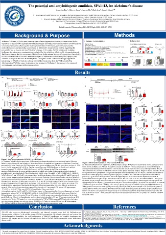
Figure 1 long -term treatment of SPA1413 in 5xFAD mice. Relative abundance(vs 5xFAD-V) 1.0 Relative abundance(vs 5xFAD-V) 1.0
(a) Treatment schedule for the experiment. (b) Body weight changes throughout the experimental period. Memory 0.0 5xFAD-V 5xFAD-SPA1413 0.0 5xFAD-V 5xFAD-SPA1413
index (c), total distance (d), and velocity (e) in the novel object recognition test. Spontaneous alterations (f) and total Figure 2 Short-term treatment of SPA1413 in 5xFAD mice.
arm entries (g) in the Y-maze test. (h) Latency time in the passive avoidance test. All data were given as means ± SEM (a) Treatment schedule for the experiment.(b) Body-weight changes throughout the experimental period. (c) Latency time
(n = 6-10 mice per group). &P < 0.05 vs. Familiar object (Two-way ANOVA followed by Bonferroni's multiple in the passive avoidance test. All data were given as means ± SEM (n = 7-9 mice per group). (d) Immunohistological
comparisons tests); *P < 0.05 vs. 5xFAD-V (One-way ANOVA followed by the Fisher exact tests). staining of amyloid plaques with thioflavin S (Thio-S) and anti-Aβ (6E10) in the cortex (CX) and hippocampus (HP) of
Amount of Aβ plaques in the cortex and hippocampus of 5xFAD mice brains. (i) Immunohistological staining of WT-V, WT-SPA1413, 5xFAD-V, and 5xFAD-SPA1413 mice groups (Scale bar 200μm). Images of the red boxes in CX and
amyloid plaques with thioflavin S (Thio-S) and anti-Aβ (6E10) in the CX and HP of WT-V, WTSPA1413, 5xFAD-V, HP regions of 5xFAD groups were enlarged and displayed in the right panel (Scale bar 100μm). Quantification analysis of
5xFAD-SPA1413, and 5xFAD-DP mice groups (Scale bar 200μm). Images of the red boxes in CX and HP regions of thioflavin S-stained plaques (e) and 6E10-positive plaques (f) numbers in CX and HP was represented as a number of
5xFAD groups were enlarged and displayed in the right panel (Scale bar 100μm). Quantification analysis of thioflavin plaques (n). (g) Activated microglia in the cortex and hippocampus of 5xFAD mice brains. Representative MHC II
S-stained plaques (j) and 6E10-positive plaques (k) numbers in CX and HP was represented as a number of plaques (n). immunofluorescent red signal images in the brains of WT-V, WT-SPA1413, 5xFAD-V, and 5xFAD-SPA1413 mice group
Oligomeric Aβ proteins (~70 kDa) were detected in CX (l) and HP (m) brain lysates of 5xFAD mice by western blot (Scale bar 200μm). Images of the red boxes in CX and HP regions of 5xFAD groups were enlarged and displayed in the
analysis. Representative full-length blots of oligomeric Aβ proteins from CX and HP brain lysates were shown. The right panel (Scale bar 100μm). (h) Quantification analysis of activated microglia in CX and HP was represented as numbers.
graph shows the percentage of the density of oligomeric Aβ proteins normalized to β-actin. All data are shown as All data were given as means ± SEM (n = 3-5 mice per group). *P < 0.05 vs. 5xFAD-V (One-way ANOVA followed by
means ± SEM, and each experiment was repeated five times (n = 4-5 per group). CX, cortex; HP, hippocampus. Tukey post-hoc comparisons tests). (i) Oligomeric Aβ proteins (60-70 kDa) were detected in CX and HP brain lysates of
*P < 0.05 vs. 5xFAD-V (One-way ANOVA followed by the Fisher exact tests). 5xFAD mice by western blot analysis. Representative full-length blots of oligomeric Aβ proteins from CX and HP brain
Activated microglia in the CX and HP of 5xFAD mice brains. (n) Representative Iba-1 immunofluorescent red signal lysates were shown. The graph shows the percentage of the density of oligomeric Aβ proteins normalized to β-actin. All
images in the brains of WT-V, WT-SPA1413, 5xFAD-V, 5xFAD-SPA1413, and 5xFAD-DP mice group (Scale bar data are shown as means ± SEM, and each experiment was repeated five times (n = 4 per group). *P < 0.05 vs. 5xFAD-V
200μm). Images of the red boxes in CX and HP regions of 5xFAD groups were enlarged and displayed in the right (Unpaired t-test).
panel (Scale bar 100μm). (o) Quantification analysis of activated microglia in CX and HP was represented as an ROI
intensity ratio (%). (p) CX lysates from mice were subjected to TNF-α ELISA assay and TNF-α levels in the brains
were investigated. All data were given as means ± SEM (n = 3-5 mice per group). CX, cortex; HP, hippocampus.
*P < 0.05 vs. 5xFAD-V (One-way ANOVA followed by Turkey post-hoc comparisons tests).
Conclusion References
SPA1413, also known as dehydroequol, phenoxodiol, and idronoxil, revealed potent both Aβ fibrilization and 1. Akiyama, H., Barger, S., Barnum, S., Bradt, B., Bauer, J., Cole, G. M., . . . Wyss-Coray, T. (2000). Inflammation and Alzheimer's disease. Neurobiol Aging, 21(3), 383-421.
doi:10.1016/s0197-4580(00)00124-x
oligomerization inhibition. In the cellular system, SPA1413 prevented the Aβ-induced cytotoxicity and reduced the 2. Gervais, F., Paquette, J., Morissette, C., Krzywkowski, P., Yu, M., Azzi, M., . . . Tremblay, P. (2007). Targeting soluble Aβ peptide with Tramiprosate for the treatment of brain
amyloidosis. Neurobiology of Aging, 28(4), 537-547. Retrieved from http://www.sciencedirect.com/science/article/pii/S0197458006000790
neuroinflammation. Remarkably, the oral administration of SPA1413 ameliorated the cognitive impairment and 3. Lovestone, S., & Smith, U. (2014). Advanced glycation end products, dementia, and diabetes. Proc Natl Acad Sci U S A, 111(13), 4743-4744. doi:10.1073/pnas.1402277111
decreased Aβ plaques and the activated microglia in the brain of 5xFAD (B6SJL) transgenic mouse Alzheimer’s 4. McLaurin, J., Kierstead, M. E., Brown, M. E., Hawkes, C. A., Lambermon, M. H. L., Phinney, A. L., . . . George-Hyslop, P. S. (2006). Cyclohexanehexol inhibitors of Aβ
disease model. aggregation prevent and reverse Alzheimer phenotype in a mouse model. Nature Medicine, 12(7), 801-808. doi:10.1038/nm1423
Acknowledgments
This work was supported by a grant from the National Research Foundation of Korea (NRF-2020R1A2B5B01002463 and NRF-2018R1D1A1B07049500) and Bio & Medical Technology Development Program of the National Research Foundation (NRF) & funded
by the Korean government (MSIT) (No. 2020M3A9E4104384). We acknowledge the assistance of Dana Kim in conducting TEM images.

