


Page 87 - ebook
P. 87
The Regulation of PCSK9 and Wnt/β-catenin Signaling
                        Pathway in Myocardial Ischemic/Reperfusion Injury

                        Yun-Ji Cha¹ , Hyoeun Kim¹ , Sae-Bom Jeon¹, Sun-Ho Lee¹², Seok-Min Kang³, Ho-Geun Yoon¹², Chan Joo Lee³,
                                ,#
                                                                ,
                                                                                        ,
                                           ,#
                                                , ,
                        Seung-Hyun Lee¹, Sahng Wook Park¹²*
                        Presenting Author : Yun-Ji Cha¹ ,#  #First Author, *Corresponding Author
                        1 Department of Biochemistry and Molecular Biology, Yonsei University College of Medicine, Seoul 03722, Korea
                        2 Graduate School of Medical Science, Brain Korea 21 PLUS Project, Yonsei University College of Medicine, Seoul 03722, Korea
                        3 Division of Cardiology, Severance Cardiovascular Hospital, Cardiovascular Research Institute, Yonsei University College of Medicine, Seoul 03722, Korea
                BACKGROUND                               Figure 3. Overexpression of Pcsk9 downregulated at the mRNA expression levels
                                                         of Wnt-related genes.
                                                         Real-time PCR data demonstrated the hypoxia-induced downregulated expression of the
   Low-density lipoprotein receptors (LDLR) play a critical role in regulating blood cholesterol levels by  Wnt/β-catenin signaling-related genes Axin2 (A) and Cyclin D1 (B). And similar to the
   binding to and clearing LDLs from circulation. The disruption of the interaction between the proprotein  previous results (Fig 1, Fig 2), the expression of Wnt/β-catenin signaling-related genes in the
   convertase subtilisin/kexin 9 (PCSK9) and LDLR reduces blood cholesterol levels. Circulating PCSK9  control group showed a tendency to recover after H/R, but did not in Pcsk9 overexpressed
   binds to the EGF domain of LDLRs causing the co-internalization of both PCSK9 and LDLR and directing  HL-1 cells. The data were normalized using Gapdh and presented as the means ± SD, by
   the LDLR to degradation in the lysosomes, rather than its recycling to the plasma membrane. Indeed,  ANOVA; n=3, *P<0.05.
   PCSK9 exerts various effects in the heart independently of LDL-cholesterol regulation. Acute myocardial
   infarction (AMI) induces local and systemic inflammation and reactive oxygen species generation,  A
   resulting in increased PCSK9 expression in cardiomyocytes. Previous study showed that PCSK9 is
   upregulated in ischemic heart tissue and determined the development of infarct size and cardiac function.  Permanent Ligation  Ischemia-reperfusion
   The Wnt/β-catenin signaling pathway plays a crucial role during vertebrate heart development, and it is
   re-activated in response to cardiac injury. In vertebrates, low-density lipoprotein receptor-related proteins
   5 (LRP5), the single-pass transmembrane protein, act as the co-receptors of Wnt ligands and is  LAD  LAD
   indispensable for Wnt signal transduction. However, It is not well known whether other members of the
   LDLR superfamily may be targets of PCSK9. The aims of this study were to determine if LDLR-related  Occlusion  Occlusion
   protein 5(LRP5) is a PCSK9 target and to study the association between Wnt/β-catenin signaling, PCSK9
   and aiming to identify possible underlying mechanisms.
                                                          Infarct      Infarct  Boarder Zone
                     RESULTS
                                                         B
    A                           C
                                           *
                                            *     *
                                          *      *
                                                         C
    B
                                             *
                                          *
                                           *
                                                * *
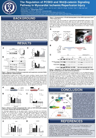
                                                         Figure 4. Regulation of Wnt/β-catenin signaling pathway in the PCSK9 cardiac-
                                                         specific overexpressed mice after ischemia-reperfusion.
   Figure 1. Hypoxia induces Pcsk9 gene expression and suppresses canonical Wnt/β-  To examine whether PCSK9 regulates injury of cardiomyocytes in vivo, we subjected transgenic
   catenin signaling in cardiomyocytes.                  mice with cardiac-specific overexpression of PCSK9 (PCSK9 Tg) and age-matched wild-type
   (A) Representative western blots of Pcsk9 under hypoxic conditions. Hypoxia induced Pcsk9  mice (WT) to ischemia-reperfusion (IR) injury induced by 1 hour of ligation of the left anterior
   expression at the protein level in HL-1 cells (B) Western blot detected the expression of Wnt  descending artery and 1 hour of reperfusion. (A),(B) Schematic illustration for the mouse
   signaling pathway key molecules. Statistically significant differences were shown that decreased  myocardial ischemia‐reperfusion model. (C) Western blot analysis of Wnt signaling pathway key
   the values of Lrp5 (0.4-fold) and phospho-β-catenin (0.5-fold) compared with baseline under  molecules in WT and hPCSK9-Tg Myh6-cre mouse hearts after 60 min of IR. α-tubulin was internal
   hypoxic conditions. After hypoxia and re-oxygenation (H/R), protein levels of Lrp5 and phospho-β-  loading control for protein levels. The results showed that 60 min of ischemia was downregulated
   catenin recovered to baseline in the control group. However, Pcsk9 overexpressed HL-1 cells, the  the Lrp5 and phospho-β-catenin compared with WT sham, similar features were observed that
   expression of phospho-β-catenin did not recover compared to control group after H/R. (C)  they are recovered after IR with in vitro results. However, sham PCSK9 Tg showed moderately
   Quantitative expression levels of proteins were shown. The data were quantified by densitometry  reduced expression levels of Lrp 5 and phospho-β-catenin compared with sham WT but did not
   and normalized to β-actin and presented as the means ± SD, by ANOVA; n=3, *P<0.05.  recover the expression of those genes after IR PCSK9 Tg compared to sham PCSK9 Tg. I,
                                                         ischemia; IR, ischemia-reperfusion.
    A
                                                                      CONCLUSION
    B                          C                                                   In this study, we investigated that LDLR-related
                                                                                   protein 5(LRP5) is a PCSK9 target, and correlation
                                                                                   with Wnt/β-catenin signaling pathway using Western
                                                                                   blot, Luciferase assay and qPCR in vitro and in vivo.
                                                                                   These results showed that PCSK9 increased under
                                                                                   hypoxic conditions and the Wnt/β-catenin signaling
                                                                                   pathway-related  genes  were  downregulated  in
                                                                                   cardiomyocytes, as demonstrated by decreased Lrp5
                                                                                   and active β-catenin protein expression levels and
                                                                                   mRNA expression levels of Axin2, Cyclin D1.
                                                                                   Interestingly, the downregulated Wnt/β-catenin-related
                                                                                   genes were recovered after H/R in the control group
                                                                                   but did not recover in the PCSK9 overexpressed
   Figure 2. Effects of Pcsk9 overexpressed HL-1 cells on TOP-/FOP-FLASH analysis  group. These results indicated that PCSK9 and Wnt/β-
                                                                                   catenin signaling were correlated at H/R conditions.
   in normoxia, hypoxia and hypoxia/re-oxygenation.                                Additionally, we found that the same results were
   (A) Schematic representation of the assay system involving TOP-Flash and FOP-Flash  obtained in the PCSK9 Tg mice. Our findings suggest
   reporters. (B) TOP-Flash and (C) FOP-Flash reporter activity determined the transcriptional  that PCSK9 and Wnt/β-catenin signaling pathway
                                                                                   could be used as a novel drug target in patients with
   activity of the β-catenin-TCF complex. (B) Reporter assays showed that hypoxia  ischemic cardiomyopathy. We need further studies to
   downregulated Wnt reporter (TOP-Flash) luciferase activity in both control and Pcsk9  evaluate the direct mechanisms of PCSK9 to the
   overexpressed HL-1 cells. The β-catenin activity was reduced under hypoxic conditions and  Wnt/β-catenin  signaling  pathway  and  obtain
   was rescued after H/R in the control group but not in Pcsk9 overexpressed cells. The values  phenotypes in human iPSC-derived cardiomyocytes.
   represent the mean ± SD, by ANOVA; n=3, *p<0.05.
    A                          B                                     REFERENCES
                                                        1. Ding Z, Wang X, Liu S, Shahanawaz J, Theus S, Fan Y, et al. PCSK9 expression in the
                                                        ischaemic heart and its relationship to infarct size, cardiac function, and development of
                                                        autophagy. Cardiovasc Res 2018;114:1738-51.
                                                        2. Badimon L, Luquero A, Crespo J, Peña E, Borrell-Pages M. PCSK9 and LRP5 in
                                                        macrophage lipid internalization and inflammation. Cardiovascular Research 2021;117:2054-68.
                                                        3. Wang X, Li X, Liu S, Brickell AN, Zhang J, Wu Z, et al. PCSK9 regulates pyroptosis via
                                                        mtDNA damage in chronic myocardial ischemia. Basic Research in Cardiology 2020;115:66.
                                                        4. Bergmann MW, Kühl M. WNT Signaling in Adult Cardiac Hypertrophy and Remodeling.
                                                        Circulation Research 2010;107:1198-208.

