


Page 83 - ebook
P. 83
Phospholipase D1 mediate flow dependent vascular inflammation

                                             Yujin Jang, Sanguk Yun*
                Department of Biotechnology, Inje University, 197 Inje-ro, Gimhae-si, Gyengsangnam-do, Korea


                         Abstract                                              Result

     PLD1 has been implicated in a variety of inflammatory diseases,
     including arthritis, asthma, and cancer. However, little is known about
     the role of PLD1 in vascular inflammation. As a result of checking the
     mRNA level using qPCR, it was confirmed that PLD1 was knocked down.
     In EC, siRNA-mediated PLD-1 gene knockdown led to the blockade of
     disturbed flow-induced increased activity of NF-kB. In addition, it was
     confirmed that siPLD knockdown in EC increased the perturbation-
     induced NF-kB activity and decreased the Yap activity. These results
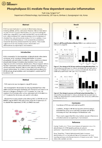
     suggest that PLD1 may mediate vascular inflammation and   Figure 1. qPCR quantification of Bovine PLD1. It was confirmed whether
                                                          PLD1 was KD using qPCR.
     atherosclerosis by regulating EC mechanization.


                       Introduction

     PLD1 is responsible for the degradation of phospholipids in the plasma
     membrane, which hydrolyzes phosphatidylcholine to produce
     phosphatidic acid and choline. In addition, various membrane-induced
     phospholipid changes themselves play important roles in vesicle
     migration and other membrane-related events. Phospholipase D 1(PLD1)
     has been implicated in various inflammatory diseases including arthritis,
                                                          Figure 2. The change of NF-kB was confirmed using disturbed flow. The
     asthma and cancer. However, little is known about the role of PLD1 in
                                                          disturbed flow was performed for 12 hours. (A) Western blot was performed to
     vascular inflammation. So, to find out how disturbed flow affects PLD,   confirm the NF-kB(p65) protein level in control and PLD KD cells. (B) p-p65 was
     NF-kB and yap phosphorylation were investigated in control and PLD   confirmed by quantification with t-p65. N=3, (**p=0.00454)
     knockdown cell.

                         Method

     - PLD1 expression was investigated using qPCR analysis.
     - We investigated NF-kB activation by inducing disturbed flow in the
     siRNA-mediated PLD1 gene knockdown cell. Fibronectin was mixed in 1X
     PBS coat in the glass, and kept in a 37℃ incubator for 1 hour, UV
     treatment for 20 minutes after suction, and coated with 0.2% dBSA in a   Figure 3. The change in yap was confirmed using disturbed flow. The
     37 ℃ incubator for 20 minutes, and used for cell culture.  disturbed flow was performed for 12 hours. (A) Western blot was performed to
                                                          confirm the Yap protein level in control and PLD KD cells. (B) Yap was confirmed by
                                                          quantification with actin.
     - All experiments were carried out by culturing BAEC in 10% FBS and p/s
     DMEM using GenDPOT’s FBS and Hyclone’s Penicillin Streptomycin. In
     the disturb flow experiment, 1% FBS, p/s DMEM was used.
                                                                             Conclusion
                                                          -  Knockdown of PLD was confirmed using qPCR.
                                                          -  In addition, changes in NF-kB and Yap were observed using in
                                                             disturbed flow using control and PLD Knockdown cell. NF-kB is
                                                             increasing and Yap is decreasing in PLD knockdown cells.
                                                          -  These results suggest that PLD1 may mediate vascular inflammation
                                                             and atherosclerosis by regulating EC mechanization.

                                                                             Reference

                                                          Huang, Chunfa, et al. "Shear stress induces cell apoptosis via a c-Src-
                                                          phospholipase D-mTOR signaling pathway in cultured
                                                          podocytes." Experimental cell research 318.10 (2012): 1075-1085.

                                                          Kang, Dong Woo. "Platelet derived growth factor increases
                                                          phospholipase D1 but not phospholipase D2 expression via NFκB
                                                          signaling pathway and enhances invasion of breast cancer cells." Cancer
                                      Created with BioRender.com  letters 294.1 (2010): 125-133.

