


Page 79 - ebook
P. 79
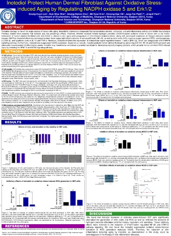
Inotodiol Protect Human Dermal Fibroblast Against Oxidative Stress-
induced Aging by Regulating NADPH oxidase 5 and Erk1/2
Seung Hoon Lee , Gun-Woo Won , Seung-Hyeon Choi , Mi-Yoon Kim , Cheong-Hae Oh , Jong-Tae Park , *, Jong-Il Park ,*
1
1
1
1
1
2 3
1
1 Department of Biochemistry, College of Medicine, Chungnam National University, Daejeon 35015, Korea
2 Department of Food Science and Technology, Chungnam National University, Daejeon 34134, Korea
3 CARBOEXPERT Inc., Daejeon 34134, Republic of Korea
ABSTRACT
Oxidative damage is one of the major causes of human skin aging. Inotodiol is a lanostane triterpenoid that demonstrates antiviral, anticancer, and anti-inflammatory activity and inhibits food allergies.
Previous studies have reported that inotodiol also has antiallergic effects. However, whether inotodiol inhibits hydrogen peroxide (H2O2)-induced oxidative stress in human skin is not known.
Stimulation of human dermal fibroblast cells with H2O2 is related to skin aging and upregulation of inflammation-related proteins, along with decreased expression of mitogen-activated protein
kinases (MAPKs). Inotodiol effectively decreased nuclear factor kappa-light-chain-enhancer of activated B cells (NF-κB), as well as nitric oxide (NO), reactive oxygen species (ROS), cyclooxygenase-
2 (COX-2), and cytokines such as IL-1β and TNF-α. Moreover, inotodiol inhibited the expression of extracellular-signal-regulated kinases 1 and 2 (ERK1/2), which are components of the MAPK
signaling pathway. Furthermore, inotodiol also suppressed NOX5 expression. Based on our results, inotodiol protects human dermal fibroblast by preventing ERK1/2, NF-κB and NOX5 activation and
attenuates the expression of inflammation genes. Inotodiol may therefore be considered a potential candidate for developing natural antiaging products, which protects human skin from ROS-induced
injury by inhibiting the ERK1/2 and NOX5 signaling pathway.
METHODS Effects of inotodiol on oxidative stress-induced inflammation in HDF cells
♣ Cell culture: Human dermal fibroblast (HDF) cells were derived from patients undergoing plastic surgery and were
provided by the Chungnam National University Hospital. The HDF cells were maintained in Dulbecco’s modified Eagle’s
medium (WelGENE, Daegu, Korea) supplemented with 10% fetal bovine serum (Gibco, maryland, USA) and antibiotics
(100U/mL penicillin and 100 μg/mL streptomycin) at 37℃ in 5% CO2. The cultured cells were stored in a nitrogen tank.
Cells between passages 4 and 9 were used in subsequent experiments. The Ethics statement was approved by the
Institutional Review Board (IRB) of Chungnam National University Hospital (IRB No: 2020-11-001) and the study was
performed according to the approved protocol.
♣ Cell viability: Cell viability was measured by using Cell Counting Kit-8 (CCK-8) assay (WST-8, Biomax, Seoul, Korea),
which detects dehydrogenase activity in the presence of an electro mediator. The HDF cells were seeded at a density of
1,000 cell/well in 96-well plate and were treated with different concentrations of hydrogen peroxide and inotodiol. And
then the plates were incubated for 24 h at 37℃ in 5% CO2. After incubation, 10 µL of CCK-8 solution was added to each
well of the 96-well plate and the wells were incubated for 1-4 hours. The absorbance at 450 nm was measured using a
microplate reader (PerkinElmer, massachusetts, USA).
♣ ROS assay : The HDF cells were homogenized in cell lysis buffer (Cell Signaling Technology, massachusetts, USA)
and centrifuged at 10,000 rpm for 15 min at 4 ℃. The supernatant was used for the quantification of ROS levels, which
was measured with DCF ROS/RNS Assay Kit (Abcam, #238535, Cambridge, UK) using dichlorodihydrofluoresc in
DiOxyQ (DCFHDiOxyQ), a fluorogenic probe specific for ROS/RNS according to the manufacturer’s instructions. The
assay is based on the principle that fluorescence intensity reflects the levels of ROS/RNS present in the sample. Intensity
was measured at excitation wavelength of 480 nm and emission wavelength of 530 nm.
Figure 3. The effects of inotodiol on oxidative stress-induced inflammation related gene in HDF cells. After serum
♣ ELISA : The NO synthesis was analyzed by determining the accumulation of nitrite (NO2-) in culture supernatant using starvation, cells were treated with Inotodiol for 4 h, and then stimulated with H O for 20 h. Quantification of cellular
2
2
a Griess Reagent System (Promega, #G2930, Wisconsin, USA). The supernatant and sulfanilamide solution were mixed inflammatory factor mRNA; IL-6, IL-17, IL-1β, TNF-α and COX-2. Statistical significance: **P <.01 compared with the
for 20 min at room temperature, then added to a N-(1-Naphthyl) ethylenediamine solution for an additional 5-10 min. The control group; ##<.01compared with the hydrogen peroxide group.
absorbance was measured at 520 nm by using a spectrophotometer (Versamax microplate reader, Molecular Device).
TNF-α (Abcam, #181421), IL-1β (R&D Systems, #SLB50, Minneapolis ,USA), ERK1/2 (Abcam, #176660), and COX-2 Effects of inotodiol on oxidative stress-induced inflammation in HDF cells
(Cell Signaling, #7291C) were measured using an ELISA kit according to the manufacture’s instructions.
♣ RNA isolation and Quantitative RT-PCR : According to the manufacturer's instructions, total RNAs from the HDF cell
was extracted using TRIzol reagent (Thermo Fisher Scientific, Massachusetts, USA). AccuPower RT PreMix (Bioneer,
Daejeon, Korea) was used for complementary DNA (cDNA) synthesis according to the manufacturer's instructions. The
obtained cDNA was amplified with specific primers (Table 1). Polymerase chain reaction (PCR) was performed for cDNA
synthesis using a T100 Thermal Cycler (Bio-Rad, California, USA). The mRNA expression was analyzed using a CFX
Connect Real-Time PCR Detection System (Bio-Rad) with PowerUp SYBR Green Master Mix (Applied Biosystems,
Massachusetts, USA). All PCR assays were performed in triplicate. The relative gene expression was analyzed using the
2−∆∆Ct method.
RESULTS Figure 4. The effects of inotodiol on oxidative stress- induced inflammation cytokines in HDF cells. After serum
starvation, cells were treated with Inotodiol for 4 h, and then stimulated with H O for 20 h. (a) TNF-α, (b) IL-1β and (c)
2
2
Effects of H O and inotodiol on the viability in HDF cells. COX-2 levels were measured by ELISA assay kit. Statistical significance: **P <.01 compared with the control group;
2
2
##<.01compared with the hydrogen peroxide group; ###<.001compared with the hydrogen peroxide group.
Inhibition effects of inotodiol on oxidative stress-NOX5 in HDF cells
Figure 5. The effects of inotodiol on oxidative stress-induced NOX5 activation in HDF cells. After serum starvation, cells
were treated with Inotodiol for 4 h, and then stimulated with H O2 for 20 h. (a) Western blot analysis was conducted to
2
2
measure NOX5. Actin was used as a loading control. (b) The graphs show the densitometric intensities of NOX5 gene
normalized to GAPDH. Statistical significance: **P <.01 compared with the control group.
Inhibition effects of inotodiol on oxidative stress-NOX5 in HDF cells
Figure 1. Cytotoxicity of H O and Inotodiol in HDF cells. (a) Chemical structure of the Inotodiol. (b) The cells were
2
2
seeded on 96-well plates and treated with H O (0-5 μM) for 24 h. Statistical significance: *P <.05 compared with the
2
2
control group. (c) The cells were seeded on 96-well plates and treated with Inotodiol (0-2 μg/mL) for 24 h. (d) The cells
were pre-treated Inotodiol (2 μg/mL) for 4 h followed by treatment with H2O2 (5 μM) for 20 h. Statistical significance: *P
<.05 compared with the control group. (e) Light microscope images. It is related to figure (d). These results are
expressed as the mean ±SEM of three independent experiment.
Inhibitory effects of inotodiol on oxidative stress-induced ROS generation in HDF cells
Figure 6. The effects of inotodiol on oxidative stress-induced p-ERK1/2 and NF-κB-p65 activation in HDF cells. After
serum starvation, cells were treated with inotodiol for 4 h, and then stimulated with H O for 20 h. (a) Western blot
2
2
analysis was conducted to measure p-ERK and NF-κB-p65. Actin was used as a loading control. (b) ERK1/2 levels
were measured by ELISA kit.
Figure 2. The effect of inotodiol on oxidative stress-induced NO and ROS generation in HDF cells. After serum DISCUSSION
starvation, cells were treated with Inotodiol for 4 h, and then stimulated with H O for 20 h. (a) The culture supernatant
2
2
was assessed using a Griess reagent system for NO generation. Statistical significance: ***P <.001 compared with the We found that inotodiol treatment of oxidative stress-induced HDF cells significantly
control group; ##<.01compared with the hydrogen peroxide group. (b) intracellular ROS levels were determined using
DCFH-DiOxyQ. DCFH-DiOxyQ fluorescence values are expressed as the fluorescence. Statistical significance: **P attenuated the generation of nitric oxide and ROS as well as inhibited the secretion of
<.005 compared with the control group. hydrogen peroxide-stimulated proinflammatory regulators and cytokines. These inhibitory
REFERENCES effects were involved to the reduction of inflammation regulated NF-κB and ERK1/2
pathway signaling. We also found that inotodiol suppressed oxidative stress-induced
Bosset S et al.Skin ageing: clinical and histopathologic study of permanent and reducible wrinkles. Eur J Dermatol. 2002, 12, 247-52. activation of ROS generation molecule NOX5. Therefore, the reduction of skin
Varani J, et al. Inhibition of type I procollagen production in photodamage: correlation between presence of high molecular weight inflammation including aging by inotodiol, as demonstrated in this study, could be
collagen fragments and reduced procollagen synthesis. J Invest Dermatol. 2002, 119, 122-129. advantageous in the therapy of skin inflammation diseases.

