


Page 33 - ebook
P. 33
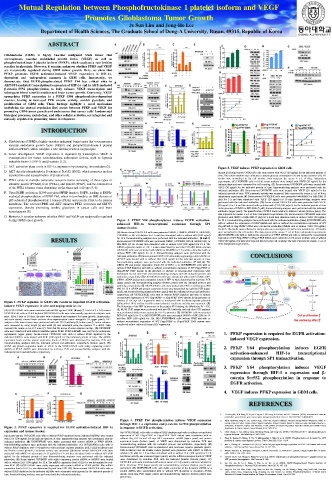
Mutual Regulation between Phosphofructokinase 1 platelet isoform and VEGF
Promotes Glioblastoma Tumor Growth
Je Sun Lim and Jong-Ho Lee
Department of Health Sciences, The Graduate School of Dong-A University, Busan, 49315, Republic of Korea
ABSTRACT
Glioblastoma (GBM) is highly vascular malignant brain tumor that
overexpresses vascular endothelial growth factor (VEGF) as well as
phosphofructokinase 1 platelet isoform (PFKP), which catalyzes a rate-limiting
reaction in glycolysis. However, it remains unknown whether PFKP and VEGF
are reciprocally regulated during GBM tumor growth. Here, we show that
PFKP promotes EGFR activation-induced VEGF expression in HIF-1α-
dependent and -independent manners in GBM cells. Importantly, we
demonstrate that EGFR-phosphorylated PFKP Y64 has critical roles in
AKT/SP1-mediated transcriptional expression of HIF-1α and in AKT-mediated
β-catenin S552 phosphorylation, to fully enhance VEGF transcription and
subsequent blood vessel formation and brain tumor growth. Conversely, VEGF
upregulates PFKP expression in a PFKP S386 phosphorylation-dependent
manner, leading to increased PFK enzyme activity, aerobic glycolysis, and
proliferation of GBM cells. These findings highlight a novel mechanism
underlying the mutual regulation that occurs between PFKP and VEGF for
promoting GBM tumor growth and underscores that cancer cells’ fundamental
biological processes, metabolism, and other cellular activities, are integrated and
mutually regulated in promoting tumor development.
INTRODUCTION
A. Glioblastoma (GBM) is highly vascular malignant brain tumor that overexpresses
vascular endothelial growth factor (VEGF) and phosphofructokinase 1 platelet
isoform (PFKP), which catalyzes a rate-limiting reaction in glycolysis.
B. tumor development. VEGF expression is regulated by transcription VEGF is
overexpressed for tumor vascularization, subsequent factors, such as hypoxia
inducible factor-1 (HIF-1) and β-catenin (1,2).
C. AKT activation plays a role in HIF-1 expression by increasing its translation (3).
Figure 5. VEGF induces PFKP expression in GBM cells.
D. AKT directly phosphorylates β-catenin at Ser552 (S552), which promotes nuclear (A and B) Serum-starved U87/EGFR cells were treated with VEGF (20 ng/ml) for the indicated periods of
translocation and transactivation of β-catenin (4). time. The culture medium was collected to analyze glucose consumption (A) and lactate secretion (B). All
results were normalized to the cell number. Data represent the means ± s.d. of three independent
E. PFK1 exists in multiple tetrameric isozymic forms consisting of three types of experiments. (C) U87/EGFR cells in 0.1% serum medium were treated with VEGF (20 ng/ml) for the
subunits: muscle (PFKM), liver (PFKL), and platelet (PFKP), and the composition indicated days and were harvested for cell counting. (D) Serum-starved U87/EGFR cells were treated with
of the PFK1 tetramer varies depending on the tissue and cell type (5, 6). VEGF (20 ng/ml) for the indicated periods of time. Immunoblotting analyses were performed with the
F. Upon EGFR activation, K395-acetylated PFKP binds to EGFR, leading to EGFR- indicated antibodies. (E) Serum-starved U87/EGFR cells were treated with VEGF (20 ng/ml) for the
indicated periods of time. PFK enzymatic activity was measured. Data represent the means ± s.d. of three
mediated phosphorylation of PFKP Y64, which in turn binds to an SH2 domain of independent experiments (F) Serum-starved U87/EGFR cells were pretreated with DMSO or MK-2206 (5
p85 subunit of phosphoinositide 3-kinases (PI3K) and recruits PI3K to the plasma μM) for 1 h and then stimulated with VEGF (20 ng/ml) for 30 min. Immunoblotting analyses were
membrane. The activated PI3K and AKT enhances PFK1 activation and GLUT1 performed with the indicated antibodies. (G) Serum-starved U87/EGFR cells were pretreated with VEGF
(20 ng/mL) for 1 h and then treated with cycloheximide (CHX;100 μg/mL) for the indicated periods of time
expression, thereby promoting aerobic glycolysis in cancer cells and brain in the presence of DMSO or MK-2206 (5 μM). Immunoblotting analyses were performed with the indicated
tumorigenesis (8). antibodies (upper panel). The quantification of PFKP levels relative to tubulin is shown (bottom panel). The
data represent the means ± s.d. of three independent experiments. (H) Serum-starved U87/EGFR cells were
G. However, it remains unknown whether PFKP and VEGF are reciprocally regulated pretreated with DMSO or MK-2206 (5 μM) for 2 h and then stimulated with or without VEGF (20 ng/mL)
during GBM tumor growth. . Figure 3. PFKP Y64 phosphorylation induces EGFR activation- for 24 h. Immunoblotting analyses were performed with the indicated antibodies. (I and J) U87/EGFR cells
enhanced HIF-1α transcriptional expression through SP1 with or without the expression of PFKP shRNA and with or without the reconstituted expression of WT
transactivation. Flag-rPFKP or Flag-rPFKP S386A were cultured in serum-free DMEM with or without VEGF (20 ng/mL)
for 48 h. The media were collected to analyze glucose consumption (I) and lactate secretion (J). All results
(A) Serum-starved U87/EGFR cells were pretreated DMSO, PD98059, SP600125, SB203580, were normalized to the cell number. The data represent the means ± s.d. of three independent experiments.
LY294002, or NF-κB inhibitor for 1 h and then stimulated with or without EGF (100 ng/ml) (K) U87/EGFR cells with or without the expression of PFKP shRNAand with or without the reconstituted
RESULTS for 12 h. Immunoblotting analyses were performed with the indicated antibodies. (B and C) expression of WT Flag-rPFKP or Flag-rPFKP S386A were cultured in 0.1% serum medium with or without
VEGF (20 ng/mL) for indicated days and harvested for cell counting. The data represent the means ± s.d. of
Serum-starved U87/EGFR cells were pretreated DMSO, LY294002, NF-κB inhibitor (B), or
MK-2206 (C) for 1h and then stimulated with or without EGF (100 ng/ml) for 12 h. The three independent experiments.
mRNA expression levels of HIF-1 α were determined by real-time PCR. (D) Serum-starved
U87/EGFR cells were pretreated with DMSO or MK2206 for 1 h and then stimulated with or
without EGF (100 ng/ml) for 12 h. Immunoblotting analyses were performed with the
indicated antibodies. (E) Serum-starved U87/EGFR cells stably expressing control shRNA or CONCLUSIONS
shPFKP were treated with or without EGF (100 ng/ml) for the indicated periods of time.
Immunoblotting analyses were performed with the indicated antibodies. (F and G) The
mRNA (F) and protein expression levels (G) of HIF-1 α in U87/EGFRvIII cells with or
without PFKP depletion and with or without reconstituted expression of WT Flag-rPFKP or
Flag-rPFKP Y64F mutant in the presence or absence of HA-myr-AKT expression were
determined by real-time PCR and immunoblotting analyses with the indicated primers and
antibodies, respectively. (H) U87/EGFRvIII cells were transfected with control siRNAor SP1
siRNA. mRNA and protein expression levels of HIF-1α were determined by real-time PCR
(upper panel) and immunoblotting analyses (bottom panel) with the indicated primers and
antibodies, respectively. (I) U87/EGFRvIII cells were treated with PBS or mithramycin (500
nM) for 12 h. mRNAand protein expression levels were determined by real-time PCR (upper
panel) and immunoblotting analyses (bottom panel) with the indicated primers and antibodies,
respectively. (J) U87/EGFRvIII cells with or without PFKP depletion and with or without
reconstituted expression of WT Flag-rPFKP or Flag-PFKP Y64F mutant in the presence or
Figure 1. PFKP depletion in GBM cells results in impaired EGFR activation- absence of HA-myr-AKT expression were co-transfected with luciferase reporter plasmids
(pGreenFire1-SP1) and the Renilla control plasmid. Luciferase activity was measured. (K)
induced VEGF expression in vitro and angiogenesis in vivo The Schematic of the HIF-1α promoter on the putative SP1 binding site (Marked as P1 – P3).
(A) U87/EGFRvIII cells were transfected with shRNA against PFKP. (B and C) Atotal of 5 × 10⁵ control (L, M, N) ChIP assays were performed with anti-SP1 antibody, and real-time PCR analyses
U87/EGFRvIII cells or PFKP-depleted U87/EGFRvIII cells were intracranially injected into athymic nude were performed with primers against the HIF-1α promoter. (L) U87/EGFR cells were treated
mice. After 5 days or 18 days, the mice were euthanized and examined for tumor growth. Hematoxylin- EGF (100 ng/ml) for 12 h. (M) U87/EGFR cells were pretreated DMOS or MK-2206 for 1h
and-eosin-stained coronal brain sections show representative tumor xenografts (B; upper panel). IHC and then EGF (100 ng/ml) for 12h. (N) U87/EGFRvIII cells without PFKP and with or
analyses of the tumor tissues were performed with anti-CD31 antibody (B; bottom panel). Tumor volumes without reconstituted expression of WT Flag-rPFKP or Flag-rPFKP Y64F mutant were
were measured by using length (a) and width (b) and calculated using the equation V = ab2/2. Data transfected with or without HA-myr-AKT expression.
represent the means ± s.d. of 5 mice (C). Note that the scores of some samples overlap. (D) U87/EGFR
cells were transfected with different shRNAs against PFKP. PFKP shRNA#1 was used for the subsequent
experiments (upper panel). Serum-starved U87/EGFR cells with or without PFKP depletion by the 1. PFKP expression is required for EGFR activation-
indicated shRNAs were treated with EGF (100 ng/ml) for the indicated periods of time. The mRNA
expression levels and the protein expression levels of VEGF were determined by real-time PCR and induced VEGF expression.
immunoblotting analyses with the indicated primers and antibodies, respectively (bottom panel). (E)
mRNA and protein expression levels of VEGF in the U87/EGFRvIII cells stably expressing control 2. PFKP Y64 phosphorylation induces EGFR
shRNA or PFKP shRNA were determined by real-time PCR and immunoblotting analyses with the
indicated primers and antibodies, respectively. activation-enhanced HIF-1α transcriptional
expression through SP1 transactivation.
3. PFKP Y64 phosphorylation induces VEGF
expression through HIF-1 α expression and β-
catenin Ser552 phosphorylation in response to
EGFR activation.
4. VEGF induces PFKPexpression in GBM cells.
REFERENCES
1. J AForsythe, B H Jiang, N V Iyer, F Agani, S W Leung, R D Koos, and G L Semenza. (1996). Activation of vascular
endothelial growth factor gene transcription by hypoxia-inducible factor 1. Mol Cell Biol 9, 4604-4613.
Figure 4. PFKP Y64 phosphorylation induces VEGF expression 2. Vijay Easwaran, Sang H. Lee, Landon Inge, Lida Guo, Cheryl Goldbeck, Evelyn Garrett, Marion Wiesmann, Pablo D.
through HIF-1 α expression and β-catenin Ser552 phosphorylation Garcia, John H. Fuller, Vivien Chan, Filippo Randazzo, Robert Gundel, Robert S. Warren, Jaime Escobedo, Sharon L.
Figure 2. PFKP expression is required for EGFR activation-induced HIF-1α in response to EGFR activation. Aukerman, Robert N. Taylor, and Wendy J. Fantl. (2003). β-Catenin Regulates Vascular Endothelial Growth Factor
Expression in Colon Cancer. Cancer Research 63, 3145-3153.
expression and transactivation. (A) U87/EGFRvIII cells with or without PFKP depletion and with or without reconstituted 3. Zhen Zhang, Li Yao, Jinhua Yang, Zhenkang Wang, and Gang Du. (2018). PI3K/Akt and HIF-1 signaling pathway in
(A) Serum-starved U87/EGFR cells with or without PFKP depletion by the indicated shRNAs were treated expression of WT Flag-rPFKP or Flag-rPFKP Y64F mutant were transfected with or hypoxia-ischemia. Mol Med Rep 4. 3457-3554.
with EGF (100 ng/ml) for the indicated periods of time. Immunoblotting analyses were performed with the without Flag-HIF-1α and HA-myr-AKT expression. mRNA (upper panel) and protein 4. Fang D, Hawke D, Zheng Y, Xia Y, Meisenhelder J, Nika H, et al. (2007). Phosphorylation of β-catenin by AKT
indicated antibodies. (B) U87/EGFRvIII were stably expressed with control shRNA or PFKP shRNA. expression levels (bottom panel) of VEGF were determined by real-time PCR and promotes β-catenin transcriptional activity. J Biol Chem 282, 11211-11219.
Immunoblotting analyses were performed with the indicated antibodies. (C) U87/EGFRvIII cells with or immunoblotting analyses with the indicated primers and antibodies, respectively. (B) 5. Mor I, Cheung EC, Vousden KH. (2011). Control of glycolysis through regulation of PFK1: old friends and recent
without PFKP depletion were co-transfected with luciferase reporter plasmids (pGL3-HRE-luciferase) and Serum-starved U87/EGFR cells co-transfected with luciferase reporter plasmids (pGL3- additions. Cold Spring Harb Symp Quant Biol 76. 211–216.
the Renilla control plasmid. Luciferase activity was measured (D) Serum-starved U87/EGFR cells were HRE-luciferase) and the Renilla control plasmid were pretreated with DMSO or HIF-1α 6. Moreno-Sanchez R, Rodriguez-Enriquez S, Marin-Hernandez A, Saavedra E. (2007). Energy metabolism in tumor
pretreated with DMSO or actinomycin D (1 μg/ml) for 1 h and then stimulated with or without EGF (100 inhibitor (10 μM) for 1 h and then stimulated with or without EGF (100 ng/ml) for 12 h. cells. FEBS J 274. 1393–1418.
ng/ml) for the indicated periods of time. Immunoblotting analyses were performed with the indicated Luciferase activity was measured (upper panel), and the mRNA expression levels of VEGF 7. Lee JH, Liu R, Li J, Zhang C, Wang Y, Cai Q, et al. (2017). Stabilization of phosphofructokinase 1 platelet isoform by
antibodies. (E) Serum-starved U87/EGFR cells stably expressing control shRNA or shPFKP were treated were determined by real-time PCR with the indicated primers (bottom panel). (C) AKT promotes tumorigenesis. Nat Commun 8. 949.
with or without EGF (100 ng/ml) for 12 h. The mRNA expression level of HIF-1α was determined by real- U87/EGFR cells were transfected with control vector or CAβ-catenin, and then cultured for 8. Lee JH, Liu R, Li J, Wang Y, Tan L, Li XJ, et al. (2018). EGFR-Phosphorylated Platelet Isoform of
time PCR. (F) U87/EGFRvIII were stably expressed with control shRNA or PFKP shRNA. The mRNA 48 h. Real-time PCR (upper panel) and immunoblotting analyses (bottom panel) were Phosphofructokinase 1 Promotes PI3K Activation. Mol Cell 70. 197–210.
expression level of HIF-1α was determined by real-time PCR. (G) Serum-starved U87/EGFR cells with or performed. (D) U87/EGFRvIII cells with stable expression of the β-catenin shRNA or a 9. Jong-Ho Lee, Fei Shao, Jinjie Ling, Sean Lu, Rui Liu, Linyong Du, Jin Woong Chung, Sang Seok Koh, Sun-Hee
without PFKP depletion by the indicated shRNAs were stimulated with hypoxia for the indicated periods of control shRNA were reconstituted with or without WT rβ-catenin or rβ-catenin S552A Leem, Jichun Shao, Dongming Xing, Zhiqiang An, and Zhimin Lu. (2020). Phosphofructokinase 1 Platelet Isofor
time. Immunoblotting analyses were performed with the indicatedantibodies. . mutant. Real-time PCR (upper panel) and immunoblotting analyses (bottom panel) were Promotes β-Catenin Transactivation for Tumor Development. Front. Oncol 10. 211.
performed.

