Page 7 - Y. Vascular biology
P. 7

Protriptyline, a tricyclic antidepressant, inhibits voltage-dependent K                                                                                                                                                                                                                                                                                                                                                                                                                +


                                                               channels in rabbit coronary arterial smooth muscle cells


                                                                                                                                                     Jin Ryeol An, Mi Seon Seo, Hee Seok Jung, Won Sun Park


                                                                   Department of Physiology, Kangwon National University School of Medicine, Chuncheon, South Korea


          Introduction


                         Tricyclic antidepressants (TCAs), including protriptyline, nortriptyline, amitriptyline clomipramine, and doxepin, are well known as the first antipsychotic agents to become widely used to


          treat depression. TCAs act by inhibiting the reuptake of two neurotransmitters, norepinephrine and serotonin, which regulate mood in the brain. TCAs are still considered effective and work well


          for many patients with depression. Of the various TCAs, protriptyline is a secondary amine TCA structurally similar to nortriptyline. Protriptyline is clinically used to treat the symptoms of


          depression and other mood disorders including anxiety. In addition, several studies reported that protriptyline improved attention-deficit hyperactivity disorder, obstructive sleep apnea, snoring,


          and tinnitus. However, apart from the useful clinical effects, protriptyline has been reported to exhibit several adverse effects including dry mouth, urinary hesitancy, confusion, ataxia, and


          constipation. The adverse effects of protriptyline on ion channels have been investigated and protriptyline has been found to block the human ether-àgo-go-related gene (hERG) potassium


          channel expressed in Xenopus oocytes and HEK293 cells. However, the cited study explored only cultured cell lines, while the side effects of protriptyline on native vascular ion channels


          [specifically voltage-dependent K (Kv) channels] have not yet been investigated.
                                                                                               +

                         Many ion channels involved in the regulation of vascular tone have been identified in vascular smooth muscle cells. Of these, K channels are the primary determinants of membrane
                                                                                                                                                                                                                                                                                                                                                                         +

          potential and constitute the predominant ion conductance pathways. Changes in membrane potential trigger opening/closing of the voltage-dependent Ca                                                                                                                                                                                                                                                                            2+      channel, thereby inducing


          vasodilation or vasoconstriction. Thus, K channels play important roles in determining vascular tone. To date, several K channels (e.g. twopore domain K channels) have been identified in
                                                                                                                                                                                                                                                                                                                                  +
                                                                                                                                                                                                                                                                                                                                                                                                                            +
                                                                                                                   +
          vascular smooth muscle cells and functional expression of four major types of K channels, including the inward rectifier K (Kir), ATP-sensitive K (K                                                                                                                                                                                                                                              ATP    ), large-conductance Ca -activated K                                                             +
                                                                                                                                                                                                                                                                                                                                      +
                                                                                                                                                                                                                         +
                                                                                                                                                                                                                                                                                                                                                                                                                                                                                 2+
                                                                                                                                                                                                                                                                                                                                                                                                  +
          (BK ), and Kv channels, has been reported. Of these, the Kv channels play a major role in suppressing membrane depolarization. Kv channel activity rises with increasing depolarization of the
                    Ca

          membrane, triggering membrane repolarization and a return to the resting membrane potential; consequently, the channels maintain the resting membrane potential and basal tone. Therefore,


          inhibition and/or overexpression of Kv channel activities may be closely associated with vascular dysfunction. In fact, several previous reports showed that inhibition of Kv channel activity


          induced membrane depolarization in mesenteric and pulmonary arterial smooth muscle cells. In addition, Kv channel expression was decreased in subjects with hypertension, hypertrophy,


          atherosclerosis, and diabetes. Thus, studies on inhibition of Kv channel activity by certain drugs are essential to prevent any unexpected effects of such drugs on vascular function.


                         In the present study, we found that protriptyline inhibited vascular Kv channels in a dose- and state-dependent manner independent of its own function.


          Material and Methods


          1. Single cell isolation Rabbit coronary arterial smooth muscle cells


          2. Patch clamp Whole cell patch clamp; Kv current


          Result


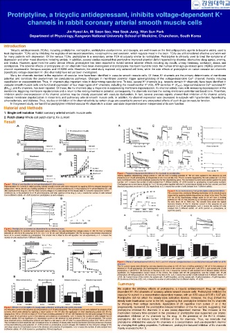
                                                                                                                                                                                                                                                                      Figure 4. Frequency-dependent inhibition of Kv channels by protriptyline.

                                                                                                                                                                                                                                                                      Twenty repetitive depolarizing voltage pulses to +60 mV from a holding potential of −80 mV were applied in the absence (o) and

                                                                                                                                                                                                                                                                      presence (•) of 10 μM protriptyline, at frequencies of (A) 1 Hz and (B) 2 Hz. The Kv currents triggered by the train pulses (at 1 or 2 Hz)

                                                                                                                                                                                                                                                                      were normalized to the current amplitude evoked by the first depolarizing pulse. The normalized currents were plotted against the pulse

                                                                                                                                                                                                                                                                      number. n=7, n means the number of cells isolated from 7 different rabbits. ∗P <0.05 (control vs. protriptyline, at each pulse number).

           Figure 1. Effect of protriptyline on voltage-dependent K (Kv) currents.                                                                                                                                                                                    Overlapping raw current traces elicited by 1 or 2 Hz train pulses together with the Kv current traces elicited by the first, second, and third
                                                                                                                    +
           Representative Kv currents in rabbit coronary arterial smooth muscle cells were measured by applying 600-ms stepped depolarizing                                                                                                                           depolarizing pulses are shown in (C,D), respectively. n=7, n means the number of cells isolated from 7 different rabbits.

           pulses (from −80 to +60 mV at a holding potential of −80 mV) in control conditions (A), in the presence of 10 μM protriptyline (B), and

           after washout of the drug (C). (D) The mean current–voltage (I–V) relationships for the steady-state Kv currents measured in control                                                                                                                                                                                                                                      Figure 5. Kv current recovery from protriptyline-induced inhibition.

           conditions (o), in the presence (•) of 10 μM protriptyline, and after washout of the drug (). n=6, n means the number of cells isolated                                                                                                                                                                                                                                   The recovery time constants following inactivation of Kv channels were

           from 6 different rabbits. ∗P <0.05 (control vs. protriptyline, at each voltage).                                                                                                                                                                                                                                                                                          measured by applying double-step pulses as shown in the inset. The first

                                                                                                                                                                                                                                                                                                                                                                                     150 ms of the depolarizing voltage pulse to 60 mV from a holding

                                                                                                                                                                                                                                                                                                                                                                                     potential of −80 mV was followed by an identical pulse with extension of

                                                                                                                                                                                                                                                                                                                                                                                     the time (from 30 to 7000 ms). The smooth lines show the typical

                                                                                                                                                                                                                                                                                                                                                                                     recovery kinetics of the Kv channel current in the absence (o) and

                                                                                                                                                                                                                                                                                                                                                                                     presence (•) of 10 μM protriptyline. n=5, n means the number of cells

                                                                                                                                                                                                                                                                                                                                                                                     isolated from 5 different rabbits. ∗P <0.05 (control vs. protriptyline, at

                                                                                                                                                                                                                                                                                                                                                                                     each time point).


           Figure 2. Protriptyline concentration-dependent Kv current responses.

           (A) Representative Kv currents were measured using a 600-ms one-step depolarizing voltage pulses of +60 mV from a holding

           potential of −80 mV in the presence of 0, 0.1, 0.3, 1, 3, 10, 30, and 100 μM protriptyline. (B) The average concentration dependence

           curve of Kv current inhibition by protriptyline. The smooth line is fitted to the Hill equation. For all experiments, n=7. n means the

           number of cells isolated from 7 different rabbits.


                                                                                                                                                                                                                                                                      Figure 6. Effects of pretreatment with Kv1.5, Kv2.1, and Kv7 channel subtype blockers on protriptyline-induced inhibition of Kv

                                                                                                                                                                                                                                                                      channels.

                                                                                                                                                                                                                                                                      The Kv current traces were elicited by one-step depolarizing pulses to +60 mV from a holding potential of −80 mV in the presence or

                                                                                                                                                                                                                                                                      absence of 10 μM protriptyline. (A) Superimposed current traces of the control, the sample with 1 μM DPO-1, and sample with 10 μM

                                                                                                                                                                                                                                                                      protriptyline +1 μM DPO-1. (B) Summary of the data of (A). n=5, n means the number of cells isolated from 5 different rabbits. NS=not

                                                                                                                                                                                                                                                                      significant. (C) Representative current traces of the control, the sample with 30 nM guangxitoxin, and the sample with 10 μM

                                                                                                                                                                                                                                                                      protriptyline +30 nM guangxitoxin. (D) Summary of the data of (C). n=5, n means the number of cells isolated from 5 different rabbits. ∗P

                                                                                                                                                                                                                                                                      <0.05. (E) Current traces of the control, the sample with 10 μM linopirdine, and the sample with 10 μM protriptyline +10 μM linopirdine.

                                                                                                                                                                                                                                                                      (F) Summary of the data of (E). n=5, n means the number of cells isolated from 5 different rabbits. ∗P <0.05.


                                                                                                                                                                                                                                                                      Summary


                                                                                                                                                                                                                                                                      We explore the inhibitory effects of protriptyline, a tricyclic antidepressant drug, on voltage-


                                                                                                                                                                                                                                                                      dependent K (Kv) channels of coronary arterial smooth muscle cells. Protriptyline inhibited the
                                                                                                                                                                                                                                                                                                      +

                                                                                                                                                                                                                                                                      vascular Kv current in a concentration-dependent manner, with an IC50 value of 5.05 ± 0.97 μM.


                                                                                                                                                                                                                                                                      Protriptyline did not affect the steady-state activation kinetics. However, the drug shifted the


                                                                                                                                                                                                                                                                      steady-state inactivation curve to the left, suggesting that protriptyline inhibited the Kv channels


                                                                                                                                                                                                                                                                      by changing their voltage sensitivity. Application of 20 repetitive train pulses (1 or 2 Hz)


           Figure 3. Effects of protriptyline on steady-state activation and inactivation of Kv channels.                                                                                                                                                             progressively increased the protriptyline-induced inhibition of the Kv current, suggesting that

           (A) Activation curves in the absence (o) and presence (•) of 10 μM protriptyline. The activation curves were obtained by analysis of tail                                                                                                                  protriptyline inhibited Kv channels in a use (state)-dependent manner. The increase in the

           currents, which were elicited by applying a return pulse of −40 mV after the application of short depolarizing pulses (20–40 ms,

           ranging from −80 to +60 mV) in steps of 10 mV from a holding potential of −80 mV. (B) Statistics for the mid-point voltage (V1/2) and                                                                                                                      inactivation recovery time constant in the presence of protriptyline also supported use (state)-

           slope value (k) of activation curve in the absence and presence of protriptyline. n=8, n means the number of cells isolated from 8                                                                                                                         dependent inhibition of Kv channels by the drug. In the presence of the Kv1.5 inhibitor,

           different rabbits. NS=not significant. (C) Inactivation curves in the absence (o) and presence (•) of 10 μM protriptyline. The curves                                                                                                                      protriptyline did not induce further inhibition of the Kv channels. Thus, we conclude that

           were obtained using a double-pulse protocol and fitted to a test step to +40 mV after 7-s preconditioning pulses from −80 to +30 mV.

           The normalized current amplitude was considered to be an estimate of steady-state channel inactivation and was plotted as a function                                                                                                                       protriptyline inhibited the vascular Kv channels in a concentration- and use-dependent manner

           of the preconditioning pulse potential. ∗P <0.05 (control vs. protriptyline, at each voltage). (D) Statistics for the mid-point voltage (V1/2)                                                                                                             by changing their gating properties. Furthermore, protriptyline-induced inhibition of Kv channels

           and slope value (k) of inactivation curve in the absence and presence of protriptyline. n=5, n means the number of cells isolated from

           5 different rabbits. ∗P <0.05 (control vs. protriptyline). NS=not significant.                                                                                                                                                                             mainly involves the Kv1.5.
   2   3   4   5   6   7   8   9   10   11   12