
Page 5 - Y. Vascular biology
P. 5
The inhibitory effect of anticholinergic drug oxybutynin on voltage-gated
K channels in coronary arterial smooth muscle cells
+
Jin Ryeol An, Won Sun Park
Department of Physiology, Kangwon National University School of Medicine, Chuncheon, South Korea
Introduction
An overactive bladder (OAB), characterized by urgency, urge incontinence and increased frequency of micturition, is associated with daily discomfort. For treatment of OAB patients,
several therapeutic drugs, such as atropine, tolterodine, darifenacin, trospium, solifenacin, fesoterodine, and oxybutynin, have been developed. The anticholinergic oxybutynin is widely used for
the treatment of symptoms of OAB, such as incontinence due to neurogenic bladder dysfunction. However, oxybutynin exerts adverse effects—such as difficulty in urination, constipation, and
delirium—in a concentration‐dependent manner. To date, however, the adverse effects of oxybutynin on vascular ion channels, specifically voltage‐dependent K (Kv) channels have not been
+
investigated.
Alterations in vascular ion channels are closely associated with many cardiovascular diseases, as well as the function of vascular smooth muscle cells. Among vascular ion channels, Kv
channels are involved in regulating membrane potential and Ca 2+ influx, which controls the contractility of vascular smooth muscle. In fact, activation of Kv channels leads to membrane
hyperpolarization, resulting in vasorelaxation, and application of a Kv channel inhibitor, such as 4‐aminopyridine (4‐AP), depolarizes the membrane potential, inducing vasoconstrictions.
Therefore, Kv channels are important therapeutic targets in several diseases; e.g., hypertension, diabetes, and hypertrophy. For example, Kv1.3 channels participate in the connection between
myocardial blood flow and cardiac metabolism. Loss of Kv1.5 subtype function is closely associated with coronary artery disease and downregulation of Kv7 subtype causes hypertension and
diabetes.
The therapeutic efficacy of oxybutynin for OAB, and the pathophysiological importance of vascular Kv channels suggest the necessity of evaluating the effect of oxybutynin on vascular Kv
channels to detect any toxic effect on the vasculature.
In this study, we investigated the inhibitory effect of oxybutynin on vascular Kv channels by using freshly isolated rabbit coronary arterial smooth muscle cells. The results showed that
oxybutynin inhibits vascular Kv channels in a concentration‐dependent manner, independent of its own function.
Material and Methods
1. Single cell isolation Rabbit coronary arterial smooth muscle cells
2. Patch clamp Whole cell patch clamp; Kv current
Result
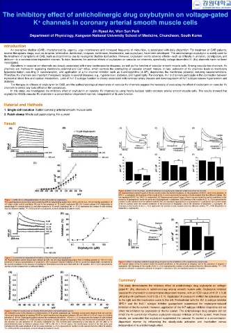
Figure 4. Effect of Kv1.5, Kv2.1, and Kv7 inhibitors on oxybutynin‐induced inhibition of the Kv current.
(A) Representative current traces of the Kv current under the control conditions, in the presence of DPO‐1, and in the presence of
DPO‐1 + oxybutynin. (B) Summary of the results of (A). n = 5 (n represents the number of cells isolated from five different rabbits). NS,
not significant (DPO‐1 vs. DPO‐1 + oxybutynin). (C) Superimposed current traces of the Kv current under the control conditions, in the
Figure 1. Inhibition of voltagedependent K (Kv) channels by oxybutynin. presence of guangxitoxin, and in the presence of guangxitoxin + oxybutynin. (D) Summary of the results of (C). n = 5 (n represents the
+
The superimposed current traces were evoked by 600‐ms depolarizing pulses from −80 to +60 mV at a −80 mV holding potential in 10 number of cells isolated from five different rabbits). NS, not significant (guangxitoxin vs guangxitoxin + oxybutynin). (E) Current traces
mV steps under control conditions (A) and in the presence of 10 μmol/L oxybutynin (B). (C) Current‐voltage (I‐V) relationship at of the Kv current under the control conditions, in the presence of linopirdine, and in the presence of linopirdine + oxybutynin. (F)
steady‐state Kv currents before (○) and after exposure to 10 μmol/L oxybutynin (●). n = 6. (n represents the number of cells isolated Summary of the results of (E). n = 5 (n represents the number of cells isolated from five different rabbits). *P < 0.05 (linopirdine vs
from six different rabbits). *P < 0.05 (control vs oxybutynin, at each voltage by Student's t‐test) linopirdine + oxybutynin)
Figure 2. Concentration‐dependent inhibition of the Kv current by oxybutynin.
(A) Representative current traces were elicited by 600‐ ms one‐step depolarizing pulses from a holding potential of −80 mV in the
presence of various concentrations of oxybutynin. (B) Oxybutynin induced inhibition of the Kv current at steady‐state, normalized to Figure 5. Effect of another anticholinergic drug atropine on the inhibition of Kv channels by oxybutynin.
the current amplitude in the absence of oxybutynin. Normalized currents were fitted with the Hill equation. All n = 6 (n represents the (A) Current traces of the Kv current under the control conditions, in the presence of atropine, and in the presence of atropine +
number of cells isolated from six different rabbits) oxybutynin. (B) Summary of the results of (A). n = 5 (n represents the number of cells isolated from five different rabbits). *P < 0.05
(control vs atropine + oxybutynin; atropine vs atropine + oxybutynin). NS, not significant (control vs atropine)
Summary
This study demonstrates the inhibitory effect of anticholinergic drug oxybutynin on voltage-
gated K (Kv) channels in rabbit coronary arterial smooth muscle cells. Oxybutynin inhibited
+
vascular Kv channels in a concentration-dependent manner, with an IC50 value of 11.51 ± 0.38
μM and a Hill coefficient (n) of 2.25 ± 0.12. Application of oxybutynin shifted the activation curve
to the right and the inactivation curve to the left. Pretreatment with the Kv1.5 subtype inhibitor
DPO-1 and the Kv2.1 subtype inhibitor guangxitoxin suppressed the oxybutynin-induced
inhibition of the Kv current. However, application of the Kv7 subtype inhibitor linopirdine did not
Figure 3. Effect of oxybutynin on steady‐state activation and inactivation curves. affect the inhibition by oxybutynin of the Kv current. The anticholinergic drug atropine did not
(A) Activation curve in the absence (○) and presence of 10 μmol/L oxybutynin (●). Activation curves were obtained from tail currents.
Tail currents were elicited by applying 30–50 ms short depolarizing step pulses between −80 and +60 mV in 10‐mV steps at a holding inhibit the Kv current nor influence oxybutynin-induced inhibition of the Kv current. From these
potential of −80 mV. Subsequent pulses returned the membrane potential to −40 mV. n = 5. (n represents the number of cells isolated results, we concluded that oxybutynin suppressed the vascular Kv current in a concentration-
from five different rabbits). *P < 0.05 (control vs oxybutynin, at each voltage by Student's t‐test). (B) Steady‐state inactivation curve in
the absence (○) and presence of 10 μmol/L oxybutynin (●). The currents were activated by a test step to +40 mV after a 7‐s dependent manner by influencing the steady-state activation and inactivation curves
preconditioning pulse of different voltages (from −80 to +30 mV in 10 mV steps). The steady‐state current of the test pulse was
normalized to the peak amplitude of the pre‐pulse potential. n = 5 (n represents the number of cells isolated from five different rabbits). independent of its anticholinergic effect.
*P < 0.05 (control vs oxybutynin, at each voltage by Student's t‐test)