
Page 5 - P. Molecular medicine and imaging
P. 5
Paeoniflorin, a Major Compound of P. lactoflora,
Increases Endometrial Receptivity by Upregulating
the Expression of LIF
Hye-Rin Park 1,2,† , Hee-Jung Choi 1,† , Mi-Ju Park , Dong-Ryeol Ryu , Ki-Tae Ha 1,2,*
1
3
Korean Medical Research Center for Healthy Aging, Pusan National University and
1
Department of Korean Medical science, School of Korean Medicine, Pusan National University, Yangsan,
2
Gyeongsangnam-do, Republic of Korea
Department of Molecular Cell Biology, Sungkyunkwan University School of Medicine, Suwon, Republic of Korea
3
These authors contributed equally in this study.
†
ABSTRACT MATERIALS & METHODS
Although recent progress of assisted reproduction technologies, many patients have been suffering from recurrent Cell viability assay: Cytotoxic effect caused by paeoniflorin
implantation failure (RIF) to achieve pregnancy after embryo transfer. Among some types of RIF, endometrial RIF treatment of cultured medium of Ishikawa cells was estimated by
defined as low endometrial receptivity and thin endometrium can be good therapeutic target of traditional herbal MTT assay.
medicine. To understand the endometrial RIF in genetic levels, we compared transcriptomic data from human Adhesion assay: The number of fluorescence-labeled JAr cells
endometrium obtained from patients undergoing RIF to normal samples. First, we performed gene set enrichment bound to Ishikawa cell monolayer were counted.
RT-PCR: The mRNA levels of LIF, several adhesion molecules
analysis using the NCBI GEO database, including GES71835, GES92324, GES26787, and GES4888. Next, we (Integrin αV, β1, β3 and β5), and β-actin were measured.
categorized functionally related gene sets with the enrichment map visualization method and the result showed the Western blot analysis: The protein levels of LIF, adhesion
cell adhesion-related gene sets were enriched in normal human endometrium. Twelve genes of them had a molecules (Integrin αV, β1, β3 and β5), and GAPDH were
positive correlation with leukemia inhibitory factor (LIF), a well-known regulator of endometrial receptivity. examined.
In our study, paeoniflorin, a major compound of P. lactoflora, enhanced embryo implantation in vitro and in vivo via Knock-down of LIF expression: pLKO.1 vectors harboring
induction of the leukemia inhibitory factor (LIF) and several integrin molecules. Therefore, our results suggest that shRNA for LIF were introduced by viral infection.
paeoniflorin might be a potent candidate for ameliorating the endometrial RIF by enhancing endometrial receptivity. In vivo study: A implantation failure model mice using RU-486,
an antagonist of progesterone receptor, were treated with
paeoniflorin(0.16mg/mouse/day).
RESULTS
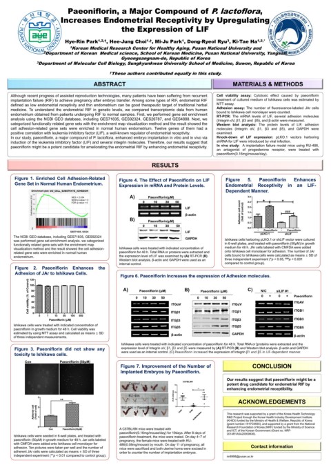
Figure 1. Enriched Cell Adhesion-Related Figure 4. The Effect of Paeoniflorin on LIF Figure 5. Paeoniflorin Enhances
Gene Set in Normal Human Endometrium. Expression in mRNA and Protein Levels. Endometrial Receptivity in an LIF-
Dependent Manner.
NES = 2.034 A) Paeoniflorin(μM) 3.0
NOM p-value = 0 * *
FDR q-value = 0 0 10 30 50 2.5
LIF 2.0
β-actin Adhesion cell count (fold of control) 1.5 ###
B) Paeoniflorin(μM) 1.0
0 10 30 50 0.5
0.0
Paeoniflorin
Paeoniflorin
GSE71835, 92324 LIF - pLKO.1 - shLIF
The NCBI GEO database, including GES71835, GES92324 GAPDH
was performed gene set enrichment analysis. we categorized Ishikawa cells harboring pLKO.1 or shLIF vector were cultured
functionally related gene sets with the enrichment map in 6-well plates, and treated with paeoniflorin (50μM) in growth
visualization method and the result showed the cell adhesion- Ishikawa cells were treated with indicated concentration of medium for 48 h. JAr cells labeled with CMFDA were added
related gene sets were enriched in normal human paeoniflorin for 48 h. Total RNA or proteins were extracted and onto Ishikawa cell monolayer for adhesion. The number of JAr
endometrium. the expression level of LIF was examined by (A) RT-PCR (B) cells bound to Ishikawa cells were calculated as means ± SD of
Western blot analysis. β-actin and GAPDH were used as an three independent experiment (*p < 0.05, ### p < 0.001
internal control. compared to control group).
Figure 2. Paeoniflorin Enhances the
Adhesion of JAr to Ishikawa Cells.
Figure 6. Paeoniflorin Increases the expression of Adhesion molecules.
120
Cell viability (% of Control)
100
A) B) C)
80 Paeoniflorin (μM) Paeoniflorin (μM) N/C siLIF #1
60 0 10 30 50 0 10 30 50 0 + 0 + Paeoniflorin
40 ITGAV
ITGαV ITGαV
20 ITGB1
0 ITGβ1 ITGβ1
0 1 10 50 100 500
Paeoniflorin (µM) ITGβ3 ITGβ3 ITGB3
Ishikawa cells were treated with indicated concentration of ITGβ5 ITGβ5
paeoniflorin in growth medium for 48 h. Cell viability was ITGB5
estimated by using MTT assay and calculated as means ± SD β-actin GAPDH
of three independent measurements. β-actin
Ishikawa cells were treated with indicated concentration of paeoniflorin for 48 h. Total RNA or ]proteins were extracted and the
Figure 3. Paeoniflorin did not show any expression level of Integrin αV, β1, β3 and β5 were measured by (A) RT-PCR (B) and Western blot analysis. β-actin and GAPDH
toxicity to Ishikawa cells. were used as an internal control. (C) Peaoniflorin increased the expression of Integrin β1 and β5 in LIF-dependent manner.
Con Paeoniflorin (50μM)
Figure 7. Improvement of the Number of CONCLUSION
Implanted Embryos by Paeoniflorin.
Our results suggest that paeoniflorin might be a
Control 15 C57BL/6N potent drug candidate for endometrial RIF by
** enhancing endometrial receptibility.
10
RU-486 # of embryos
3
** 5 ACKNOWLEDGEMENTS
Adhered cell (Fold of cell) 2 RU-486 + Paeoniflorin 0 C o n trol RU -4 8 6 R U -48 6+ P ae o niflo rin This research was supported by a grant of the Korea Health Technology
R&D Project through the Korea Health Industry Development Institute
1
(KHIDI) funded by the Ministry of Health & Welfare, Republic of Korea
(grant number: HI17C0935), and supported by a grant from the National
0 Research Foundation of Korea (NRF) funded by the Ministry of Science
Con Paeoniflorin(50μM) A C57BL/6N mice were treated with and ICT, of the Korean Government (Grant no. NRF-
paeoniflorin(0.16mg/mouse/day) for 19days. After 8 days of 2014R1A5A20009936).
Ishikawa cells were seeded in 6-well plates, and treated with paeoniflorin treatment, the mice were mated. On day 4~7 of
paeoniflorin (50μM) in growth medium for 48 h. Jar cells labeled pregnancy, the female mice were treated with RU-
with CMFDA were added onto Ishikawa cell monolayer for 486(0.08mg/mouse) by mouth. On day 11 of pregnancy, all
adhesion. Ten pictures were taken per well and the number of mice were sacrificed and both uterine horns were excised in Contact information
adherent JAr cells were calculated as means ± SD of three order to counter the number of implantation embryos.
independent experiment (**p < 0.01 compared to control group). rin8998@pusan.ac.kr