Page 7 - P. Molecular medicine and imaging
P. 7

Protein Nanobio Lab sat UNIST


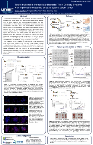
                                                                                  Target-switchable Intracellular Bacterial Toxin Delivery Systems


                                                                                  with improved therapeutic efficacy against target tumor


                                                                                  Seong Guk Park, Bongseo Choi, Yoonji Bae, Sebyung Kang


                                                                               Department of Biological Sciences, School of Life Sciences, Ulsan National Institute of Science and Technology (UNIST), Ulsan, 44919, Korea


                                                                                                                Abstract                                                                                                                                                                                                                                                    Scheme


                         Targeted cancer therapies have been extensively developed to selectively


                    suppress tumor growth as well as to avoid harming healthy tissues. However,


                    failure to escape endosome upon receptor-mediated endocytosis is a major


                    obstacle limiting the efficacy of protein-based targeted cancer therapeutics, such


                    as recombinant immunotoxin. Here, novel target-switchable intracellular toxin


                    delivery systems (TiTDS) is presented, using catalytic and translocation domain of


                    diphtheria toxin (dtA-T) as an intracellular toxin delivery platform and affibody


                    molecules targeting HER2 or EGFR (HER2Afb or EGFRAfb) as target-specific


                    ligands. The intracellular toxin delivery platform and affibody molecules are


                    genetically fused with SpyCatcher (SC) protein and SpyTag (ST) peptide,                                                                                                                                                                                         Schematic illustration of the construction of target-switchable intracellular bacteria


                    respectively, to generate dtA-T-SC and ST-HER2Afb or ST-EGFRAfb modules.                                                                                                                                                                                   toxin delivery systems and TiTDS-mediated selective cancer treatments. Toxin
                                                                                                                                                                                                                                                                               delivery module (dtA-T-SC) composed of diphtheria toxin A-T domain and SC can be


                    These modules can be individually purified and post-translationally ligated to form                                                                                                                                                                        ligated with ST-fused targeting ligands (ST-HER2Afb and ST-EGFRAfb) in a mixing-


                    dtA-T/HER2Afb or dtA-T/EGFRAfb, through simple mixing-and-matching.                                                                                                                                                                                        and-matching manner, forming dtA-T/HER2Afb or dtA-T/EGFRAfb. dtA-T/HER2Afb


                         dtA-T /HER2Afb and dtA-T/EGFRAfb can selectively bind to their corresponding                                                                                                                                                                          and dtA-T/EGFRAfb selectively bind to their corresponding target cancer cells, enter
                                                                                                                                                                                                                                                                               the cells through receptor-mediated endocytosis, and successfully release active


                    target cancer cells, efficiently enter the cells through receptor-mediated                                                                                                                                                                                 toxins to cytosol, leading to death of target cancer cells.


                    endocytosis, successfully escape endosome, and release active toxins to the


                    cytosol. They exhibit high target-specific cytotoxicity in vitro and can reduce tumor                                                                                                                                                                                                                  Target-specific toxicity of TiTDS


                    masses significantly in vivo. The TiTDS will be promising targeted cancer


                    therapeutic platforms because of their high target specificity, effective intracellular


                    delivery of active toxins with improved therapeutic efficacy, and versatile target


                    switchability.


                                                                                                Characterization


                                                                                                                                                                                                                                                                              Figure 3. Target cell-specific cytotoxicity tests with dtA-T/Afb. Time-dependent


                                                                                                                                                                                                                                                                              morphological changes of (A) NIH3T6.7 and (B) A431 cells upon treatment with PBS,


                       Figure 1. Construction and characterization of intracellular toxin delivery                                                                                                                                                                            20 μM of doxorubicin, and 2 μM of corresponding dtA-T-SC, ST-Afb, and dtA-T/Afb


                       modules (dtA-T-SC), targeting ligands (ST-Afb), and their ligated forms (dtA-                                                                                                                                                                          monitored                        by           the            real-time                    microscope,                            IncuCyte® .                         In         vitro             quantitative


                       T/Afb). (A) Schematic illustration of dtA-T-SC and ST-Afb (ST-HER2Afb and ST-                                                                                                                                                                          measurement of dose-dependent cytotoxicity of dtA-T/HER2Afb to NIH3T6.7 cells


                       EGFRAfb) constructs. L represents a linker peptide. (B) The SDS-PAGE                                                                                                                                                                                   and dtA-T/EGFRAfb to A431 cells assayed by CCK-8.


                       analysis of dtA-T-SC, ST-Afb, and dtA-T/Afb. (C) Molecular mass analyses of


                       dtA-T-SC, ST-Afb, and dtA-T/Afb with mass spectrometry. Calculated (Calc.) and


                       observed (Obs.) masses are indicated.


                                                                                                                                                                 Figure 2. Trafficking released


                                                                                                                                                                 dta upon target-cell binding of


                                                                                                                                                                 dta-T/Afb and subsequent


                                                                                                                                                                 receptor-mediated endocytosis.


                                                                                                                                                                 Confocal fluorescence


                                                                                                                                                                 microscopic cell images of (A)


                                                                                                                                                                 NIH3T6.7 cells treated with


                                                                                                                                                                 dta-T/HER2Afb and (B) A431


                                                                                                                                                                 cells with dta-T/EGFRAfb were


                                                                                                                                                                 obtained at indicated times.


                                                                                                                                                                 Inactive diphtheria toxin A


                                                                                                                                                                 fragments (dta, green) are


                                                                                                                                                                 probed with anti- diphtheria


                                                                                                                                                                 toxin A antibodies, which


                                                                                                                                                                 selectively bind to the released


                                                                                                                                                                 form of A fragment. Nuclei of


                                                                                                                                                                 cells (blue) and endosomes


                                                                                                                                                                 (red) were stained with DAPI


                                                                                                                                                                 and lysotracker, respectively.                                                                                Figure 4. Improved in vivo therapeutic efficacy of albumin-binding domain (AlBD)-


                                                                                                                                                                 Scale bar = 20 μm.
                                                                                                                                                                                                                                                                               fused dtA-T/Afb. (A) Schematic illustration of AlBD-fused dtA-T/Afb constructs and


                                                                                                                                                                                                                                                                               their expected forms after ligation with ST-Afb. (B) PBS, AlBD-fused dtA-


                                                                                                                                                                                                                                                                               T/HER2Afbs, and dtA-T/HER2Afb (5 μg, respectively) were intravenously injected to


                                                                                                                                                                                                                                                                               NIH3T6.7-allografted nude mice, and their tumor volumes were measured with a


                                                                                                                                                                                                                                                                               caliper. (C) A431 cells were inoculated to nude mice. PBS, AlBD-fused dtA-


                                                                                                                                                                                                                                                                               T/EGFRAfb, and dtA-T/EGFRAfb (5 μg, each) were intravenously injected. A total of


                                                                                                                                                                                                                                                                               eight injections at 2- to 3-day intervals were administered. Pictures of biopsied


                                                                                                                                                                                                                                                                               tumors from sacrificed mice are presented below (B) and (C).


                                                                                           Acknowledgement                                                                                                                                                                                                                                                            Conclusion


                                                                                                                                                                 Laboratory Members

                                                                                                                                                                                                                                                                                     In conclusion, our target-switchable intracellular bacterial toxin delivery system

                                                                                                                                                Ph.D. : Bongseo Choi (previous)                                                                                                    provides a great potential for developing versatile anti-cancer platforms that can


                                                                                                                                                MS/Ph.D. Candidate: Hansol Kim                                                                                                     be applied to various target cancers by adopting multiple targeting ligands on


                                                                                                                                                MS/Ph.D. Candidate: Hyukjun Choi


                                                                                                                                                MS/Ph.D. Candidate: Yoonji Bae                                                                                                     demand.
   2   3   4   5   6   7   8