
Page 27 - O. Microbiology
P. 27
(O-22) Comparative microbiomes of tonsil and saliva in pediatric patients
subjected to tonsillectomy
1
1
1
1
2
3,4
3,4
2
Da Hyeon Choi , Jiwon Park , Ju Kwang Choi , Kyeong Eun Lee , Won Hee Lee , Jinho Yang , Ju Yeon Lee , Yoon Jeong Park ,
5
Chan Oh , Ho-Ryun Won , Bon Seok Koo , Jae Won Chang 5,* Yoon Shin Park 1,*
5
5
1 Department of Microbiology, School of Biological Sciences, College of Natural Sciences, ChungbukNational University, Cheongju 28644, Republic of Korea
2Institute of MD Healthcare Inc, Seoul, Republic of Korea
3Central Research Institute, Nano Intelligent Biomedical Engineering Corporation (NIBEC), School of Dentistry, Seoul National University, Seoul 03080, Republic of Korea
4Department of Dental Regenerative Bioengineering and Dental Research Institute, School of Dentistry, Seoul National University, Seoul 03080, Republic of Korea
5Department of Otolaryngology-Head and Neck Surgery, Chungnam National University College of Medicine, Daejeon 35015, Republic of Korea
pys@cbnu.ac.kr
BACKGROUND AIM
Oral bacteria travel throughout the body and are significantly associated with human diseases.
Given that tonsils are located between the oral cavity and larynx esophagus at the gateway of
both alimentary and respiratory tracts, tonsillar tissue may also be affected by both oral and
alimentary tract microbiota. Here, we focused on the distribution and correlations of microbiota in
saliva and tonsillar tissues of young tonsillectomy patients based on evaluation of the V3-V5
region of 16S rRNA genes to examine the hypothesis that the microbiome is associated with
tonsillar hyperplasia in children. The top 10 ranked taxa in saliva group and tonsillar group
based on average relative abundance were Haemophilus, Streptococcus, Fusobacterium,
Veillonella, Prevotella, Alloprevotella, Neisseria, Prophyromonas, Campylobacter, and
Treponema 2. Analysis of the microbiomes between tonsil and saliva revealed that many
bacterial communities are shared and show similarities in terms of diversity and composition,
suggesting close interactions between the two microbial groups. Our results assume that the oral microbiome exerts significant effects on not only the tonsil
itself but also tonsil-derived immune or stem cells through regulating the microbial community.
METHODS
Sample collection: Among the 45 participants who underwent tonsillectomy at Chungnam National University Hospital (CNUH, Daejeon, Korea) between June 2018 and January 2019, 29 were
enrolled for analysis (IRB No. 2018-06-021-002).
Anthropometric measurements and serum biochemical indices of subjects: We collected anthropometric data from all subjects, including the height and weight of all patients, which were
measured the day before surgery.
DNA extraction and amplicon sequencing: DNA from saliva and tonsillar tissue was extracted using a DNeasy PowerSoil kit (Qiagen, Germany). DNA in each sample was quantified using
QIAxpert (Qiagen). Specific V3-V4 hypervariable regions of the 16S rRNA gene were amplified using 16S_V3_F (5′-TCGTCGGCAGCGTCAGATGTGTATAAGAGACAGCCTACGG
GNGGCWGCAG-3′) and 16S_V4_R (5′-GTCTCGTGGGCTCGGAGATGTGTATA AGAGACAGGACTACHVGGGTATCTAATCC-3′) primers. Libraries were prepared using PCR products and
quantified with the aid of the QIAxpert kit (Qiagen). Each amplicon was quantified, pooled, and sequenced using MiSeq (Illumina, USA). All raw sequences derived from this experiment were
submitted to the Short Read Archive of NCBI and can be found under BioProject accession number #PRJNA615768.
Analysis of bacterial compositions of microbiomes: A total of 726,274 raw reads were generated from the saliva and tonsillar tissue of 29 participants, with an average of 12,745 reads
(standard deviation 3,859). Paired-end reads matching the adapter sequences were trimmed using cutadapt (version 1.1.6). The resulting FASTQ files containing paired-end reads were merged
with CASPER and quality-filtered with a Phred (Q) score of 2017. Any reads < 350 bp or > 550 bp after merging were additionally excluded.
RESULTS
Fig.1 Microbiota compositions of saliva and tonsil samples from 29 Fig.3 Comparison of saliva and tonsil groups at the genus level
participants.
Relative abundance at the (a) phylum level and (b) genus level from 29 participants.
Relative abundance of the microbial community based on the dominant phyla and Principal coordinate analysis plots of Bray-Curtis-computed distances between saliva and tonsils.
genera. The highly abundant phyla were Firmicutes, Proteobacteria, Bacteroidetes, (a) Saliva (red) and tonsil (blue) samples are colored according to subject. The plot presents a 95%
Actinobacteria, and Fusobacteria. The predominant genera were Haemophilus, confidence ellipse with a color background. (b) Venn diagram showing overall overlap between
Fusobacterium, Streptococcus, Prevotella and Veillonella. saliva and tonsils at the genus level. (c) Number of specific and common genera between saliva
and tonsil samples at the phylum level.
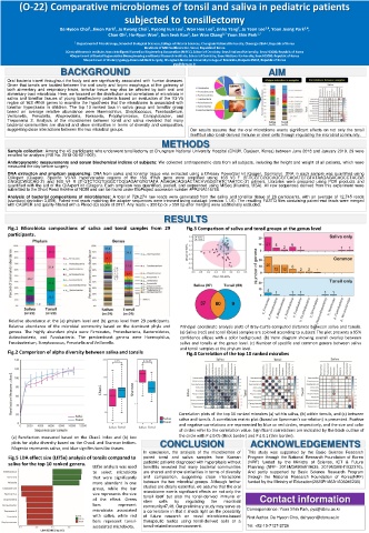
Fig.2 Comparison of alpha diversity between saliva and tonsils Fig.4 Correlation of the top 10 ranked microbes
Correlation plots of the top 10 ranked microbes (a) within saliva, (b) within tonsils, and (c) between
saliva and tonsils. A correlation matrix plot (based on Spearman’s correlation) is presented. Positive
and negative correlations are represented by blue or red circles, respectively, and the size and color
of circles refer to the correlation value. Significant correlations are indicated by the black outline of
(a) Rarefaction measured based on the Chao1 index and (b) box the circle with P ≤ 0.05 (thick border) and P ≤ 0.1 (thin border).
plots for alpha diversity based on the Chao1 and Shannon indices. CONCLUSION ACKNOWLEDGEMENTS
Magenta represents saliva, and blue signifies tonsillar tissues.
In conclusion, the analysis of the microbiomes of This study was supported by the Basic Science Research
Fig.5 LDA effect size (LEfSe) analysis of tonsils compared to paired tonsil and saliva samples from Korean Program through the National Research Foundation of Korea
saliva for the top 10 ranked genera. pediatric patients diagnosed with hyperplasia without (NRF) funded by the Ministry of Science, ICT & Future
LEfSe analysis was used tonsillitis revealed that many bacterial communities Planning (NRF- 2016M3A9B4919639, 2019M3A9H1032376).
to select microbiota are shared and show similarities in terms of diversity And partly supported by Basic Science Research Program
that were significantly and composition, suggesting close interactions through the National Research Foundation of Korea(NRF)
more abundant in one between the two microbial groups. Although further funded by the Ministry of Education(2020R1A6A1A06046235)
group, while the bar studies are clearly essential, we assume that the oral
microbiome exerts significant effects on not only the
size represents the size tonsil itself but also the tonsil-derived immune or
of the effect. Green stem cells by regulating the microbial Contact information
bars represent community47,48. Our preliminary study may serve as
microbiota associated a cornerstone in that it sheds light on the possibility Correspondence: Yoon Shin Park, pys@cbnu.ac.kr
with saliva, while red of future research on novel microbiome-based First Author: Da Hyeon Choi, dahyeon@cbnu.ac.kr
bars represent tonsil- therapeutic tactics using tonsil-derived cells or a
associated microbiota. tonsil-related microenvironment. Tel: +82-10-7127-2726