
Page 5 - J. Chromatin remodeling and epigenetics
P. 5
Lysine-specific demethylase 3A is important for autophagic occurrence
Jisu Park, Minsol Jeon, Hyunkyung Kim
Department of Biochemistry and Molecular Biology, Korea University College of Medicine, Seoul 02841, Republic of Korea
ABSTRACT
Autophagy is an essential process to maintain cell survival and homeostasis under various stress conditions. Here, we report that lysine-specific demethylase
3A (KDM3A) plays an important role in starvation-induced autophagy. Using Kdm3a knockout mice, we demonstrate that KDM3A is crucial for proper hepatic
autophagy in vivo. Hepatic mRNA expression analysis and ChIP assay in WT and Kdm3a knockout mouse livers reveal that KDM3A activates autophagy genes
by reducing histone H3K9me2 levels upon fasting. Together, our finding represents previously unidentified function of KDM3A as a key regulator of
autophagy, implicating potential therapeutic approaches for autophagy-related diseases.
INTRODUCTION
Autophagy works moderately as a basal state and can be further induced by various signals, including nutrient starvation [1]. The acute and rapid response of
autophagy mainly occurs in the cytoplasm, but recent accumulating evidence has highlighted that prolonged starvation triggers transcriptional and
epigenetic regulatory programs in the autophagic process [2]. Histone methylation and demethylation are associated with these transcriptional modulations
of autophagy and lysosomal genes [3.4]. KDM3A, a member of Jumonji domain-containing protein, is explicitly known to demethylate H3K9me1 and
H3K9me2 and requires Fe (II) and -ketoglutarate for catalytic activity, which functions as a transcriptional coactivator [5]. Here, we provide a functional link
between transcriptional regulation of autophagy and histone demethylation. KDM3A is induced upon glucose starvation and removes H3K9me2 for
transcriptional activation of autophagy genes. Further, we applied Kdm3a KO mouse model to understand how KDM3A functions as a crucial player for
proper hepatic autophagy in vivo.
RESULTS
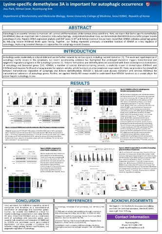
Figure 2. KDM3A is critical for autophagosome
formation and lysosomal function
(A) GFP-LC3 was transfected in WT and Kdm3a KO MEFs
and the formation of GFP-LC3 punctation was examined
by confocal microscopy. GFP-LC3 (green); DAPI (blue).
Graphs show quantification of LC3-positive punctate
cells. Values are expressed as mean ± s.d. of three
independent experiments. ***p<0.001. Scale bar,
10 μm. (B) GFP-LAMP1 was transfected in WT MEFs and
Kdm3a KO MEFs, and the activation of lysosome by
increased GFP-LAMP1 intensity was examined by
confocal microscopy. GFP-LAMP1 (green); DAPI (blue).
Graphs show quantification of intensity of LAMP1
(right). Values are expressed as mean ± s.d. of three
independent experiments. ***p<0.001. Scale bar,
10 μm. (C) WT MEFs and Kdm3a KO MEFs were
deprived of glucose for 12 hours in the absence or
presence of bafilomycin A1 (BafA1; 50 nM, 2 hours).
Autophagic flux was analyzed with anti-LC3 antibody.
The LC3-II/-actin ratio is indicated. (D) mCherry-GFP-
Figure 1. Increased KDM3A levels upon starvation are critical for proper autophagic occurrence LC3 was transfected in WT MEFs and Kdm3a KO MEFs
and the autophagosomes (mCherry-positive, GFP-
(A and B) Quantitative RT-PCR (qRT-PCR) analysis of KDM3A mRNA levels in MEFs and HepG2 positive puncta) and autolysosome (mCherry-positive,
cells in response to glucose starvation (A) or amino acid starvation (B). Values are expressed as GFP-negative puncta) formation was examined by
mean ± s.d. of three independent experiments. ***p<0.001. Statistics by one-tailed t-test. (C and confocal microscopy. DAPI (blue). Graphs show
D) Cell lysates of WT MEFs and Kdm3a KO MEFs, starved for glucose (C) or amino acid (D) for quantification of autophagosome and autolysosome
indicated times were subject to immunoblot analysis with anti-LC3 antibody. The LC3-II/-actin punctate cells. Values are expressed as mean ± s.d. of
ratio is indicated. *: non-specific band. three independent experiments. ***p<0.001. Scale bar,
10 μm.
Figure 3. KDM3A is important for Figure 4. KDM3A is responsible for activation of autophagy
proper hepatic autophagy in vivo genes in the livers of fasted mice
(A) WT and Kdm3a KO mice were fed or fasted for 24 hours
(A) Liver tissues from fed or fasted mice and mouse liver tissues were prepared for analysis. mRNA
were subject to immunoblot analysis. levels of autophagy and lysosomal genes were determined by
The LC3-II/-actin ratio is indicated. (B) qRT-PCR analysis. Each mRNA level was normalized by 36B4
Liver sections collected from WT mice gene. Values are expressed as mean ± s.d. of three
and Kdm3a KO mice upon starvation for independent experiments. ***p<0.001. Statistics by two-
24 hours were stained and compared tailed t-test. (B) Recruitment of KDM3A with decrease of
with ad libitum. Representative H3K9me2 was analyzed by ChIP assays in liver tissues from
confocal image of LC3 puncta formation fasted mice compared to fed mice. The decrease of H3K9me2
was analyzed by anti-LC3 antibody. was compromised in Kdm3a KO liver in response to starvation.
Graph shows quantification of LC3- *p<0.05, **p<0.01, ***p<0.001. (C) Schematics for KDM3A
positive punctate cells. Scale bar, 10 μm. function in regulation of autophagy.
c
Glucose
starvation
CONCLUSION REFERENCES ACKNOWLEDGEMENTS
Here, we report that KDM3A is induced by nutrient We thank H.-J.R.S. for helpful discussion and editing
starvation and functions as a transcriptional [1] Autophagy: renovation of cells and tissues, Cell. 147 (2011) 728- and S.H.K. for technical assistance. Kdm3a KO MEFs
741,
coactivator of autophagy and lysosomal genes.
Further, we validate that KDM3A is critical for [2] TFEB controls cellular lipid metabolism through a starvation- were a gift from Yi Zhang.
hepatic autophagy occurrence in vivo using Kdm3a induced autoregulatory loop, Nat Cell Biol. 15 (2013) 647-658,
KO mice. Given that H3K9me2 acts as a repressive [3] Epigenetic Control of Autophagy: Nuclear Events Gain More Contact information
mark of autophagy and lysosomal genes upon Attention, Mol Cell. 65 (2017) 781-785,
nutrient rich conditions and reduction of H3K9me2 [4] Lateral Thinking: How Histone Modifications Regulate Gene
by KDM3A are prominent event upon nutrient Expression, Trends Genet. 32 (2016) 42-56, Hyunkyung Kim,
deprivation, our data provide a functional link Phone: 82-2-2286-1299
between energy sensing, histone modifications, and [5] JHDM2A, a JmjC-containing H3K9 demethylase, facilitates
transcription activation by androgen receptor, Cell. 125 (2006) 483-
epigenetic regulation of autophagy 495 email: hyunkkim@korea.ac.kr