
Page 73 - I. Chemical biology and drug discovery
P. 73
Phenotypic discovery of a neuroprotective agent
regulating tau proteostasis in a stress-responsive
manner
a
Young-Hee Shin , Hana Cho , Jonghoon Kim , Jaeyoung Ha , Seung Bum Park* a
a
a
a
a Department of Chemistry, Department of Biophysics and Chemical Biology, Seoul National University, Seoul 08826, South Korea. Seoul National University
Abstract
A number of neurodegenerative diseases including tauopathies are caused by an abnormal proteostasis and accumulation of aggregation-prone proteins in neurons. In order to find small
molecules suppressing tau protein aggregation in cells under endoplasmic reticulum (ER) stress conditions, we performed a phenotype-based screening using HEK293 Tau BiFC (Bimolecular
Fluorescence Complementation)-Venus cells. SB1617 was selected as a leading compound via a structure activity relationship study for its high potency in reducing tau protein oligomerization and
low cytotoxicity. We obtained a possible target protein list of SB1617 by applying label-free target identification technology, TS-FITGE (thermal shift Fluorescence difference in two-dimensional gel
electrophoresis). Further gene knockdown experiments for those proteins, biophysical tests and in vitro bio-functional tests revealed that SB1617 activates protein kinase-like endoplasmic reticulum
kinase (PERK) signaling under the cell stressed conditions. Further elucidation of mechanism regarding conditional PERK activation and proteostasis regulation will accelerate developing
therapeutics for neurodegenerative diseases.
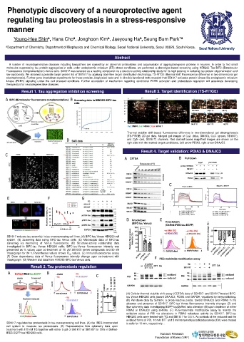
Result 1. Tau aggregation inhibition screening Result 3. Target identification (TS-FITGE)
A BiFC (Bimolecular fluorescence complementation) B Screening data in HEK293-BIFC-tau
cells
Cy2: DMSO, Cy3: SB1607, Cy5: SB1617
Thermal stability shift-based fluorescence difference in two-dimensional gel electrophoresis
(TS-FITGE) 2D gel data. Merged gel images of Cy2- (blue, DMSO), Cy3- (green, SB1607),
and Cy5- (red, SB1617) channels. Red dashed boxes magnified images are shown on the
D SAR data
right side with the marked target candidates. Left arrow-PDIA3, right arrow-DNAJC3.
Result 4. Target validation: PDIA3 & DNAJC3
C DMSO TG TG/SB1617
A CETSA B Pull down
C
E F G
DMSO TG TG/SB1617 120
Fluorescence Intensity (%) 100 80 60 SB1617 D Knockdown E Knockdown
SB1607
40
20
0.1 IC 50 1.87 0.59 M 10 100 (BiFC-tau Venus) (DsRed-IRES-tau:EGFP)
1
[SB16xx], M
SB1617 reduces tau assembly in tau-overexpressing cell lines. (A) BiFC-tau Venus HEK293 cell
system. (B) Screening data using BiFC-tau Venus cells. (C) Microscopic data of BiFC-tau
screening via monitoring of Venus fluorescence. (D) Structure-activity relationship data
investigated in BiFC-tau Venus HEK293 cellls. BiFC-tau-Venus fluorescence intensity was
presented as % values upon co-treatment of 10 μM SB16XX series compounds and 80 nM
thapsigargin for 24 h. Parentheses values shows IC 50 values. (E) Immunofluorescence assay.
(F) Dose dependency data of Venus fluorescence intensity change upon co-treatment with F
thapsigargin. (G) Western blot data from HEK293-BiFC-tau-Venus cells. PEG-maleimide modification assay
Result 2. Tau proteostasis regulation
A DsRed-IRES-tau EGFP B
C
(A) Cellular thermal stability shift assay (CETSA) data of SB1607- and SB1617-treated BiFC-
tau Venus HEK293 cells toward DNAJC3, PDIA3 and GAPDH, visualized by immunoblotting.
(B) Pull-down data by SB1624, a photo-reactive probe, toward DNAJC3 and PDIA3 in the
absence and presence of SB1617. BiFC-tau Venus fluorescence intensity changes (D) and
flow cytometry data investigating EGFP-tau/DsRed ratio alteration (E) upon depletion of either
PDIA3 or DNAJC3 using siRNAs. (F) PEG-maleimide modification assay to monitor the
oxidation status of PDI via alterations in PDIA3 reductase activity by SB1617. BiFC-tau
HEK293 cells were treated with TG and SB1617 for 3.5 h. As controls of the reduced and the
oxidized forms of PDI, 10 mM DTT and 5 mM tetramethylazodicarboxamide (DA) were treated
SB1617 regulates tau proteostasis in tau-overexpressing cell lines. (A) the IRES-incorporated to cells for 15 min, respectively. .
cell system to measure tau proteostasis. (B) Representative flow cytometry data upon
treatment with 100 nM TG together with either 5 µM of SB1617 or SB1607 for 20 h in DsRed-
IRES-EGFP-tau HEK293 cells. National Research
Foundation of Korea (NRF)