
Page 49 - I. Chemical biology and drug discovery
P. 49
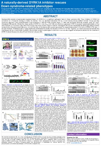
A naturally-derived DYRK1A inhibitor rescues
  Down syndrome-related phenotypes
  Young-wook Ham 1,2,# , Miri Choi 1,2,# , Ae-kyeong Kim , Joo-Youn Lee , Jung-Nyoung Heo , Min-Hyo Ki , Sang-Bae Han , Kyu-Sun Lee , Sungchan Cho 1,7,*
                                                                     6
                                                                               2
                                                                                         3
                                    3
                                               4
                                                            5
  1 Natural Medicine Research Center, KRIBB, Korea.  2 College of Pharmacy, Chungbuk National University, Korea.  3 Bionanotechnology Research Center, KRIBB, Korea.  4 Chemical Data-Driven
  Research Center, KRICT, Korea.  5 Bioorganic Science Division, KRICT, Korea.  6 Center Research Institute, Samjin Pharm. CO.,LTD, Korea. 7 Department of Biomolecular Science, KRIBB School of
  Bioscience, KUST, Korea.
                                               ABSTRACT
   Dual-specificity tyrosine phosphorylation-regulated kinase 1A (DYRK1A) is a significant pathogenic factor in Down syndrome (DS). Thus, inhibition of DYRK1A is
   considered as a therapeutic strategy to modify the disease. Here, we identified Aristolactam BIII as a novel naturally-derived DYRK1A inhibitor through two-step
   screening approach using structure-based virtual screening of >400,000 KCB chemical library (1 st  step) and cell-based NFAT-RE promoter assay (2 nd  step).
   Aristolactam BIII potently inhibited the kinase activity of DYRK1A in vitro (IC 50 = 9.67 nM) and effectively suppressed DYRK1A-mediated hyperphosphorylation of Tau
   and Presenilin 1 in mammalian cells, which was stronger than those of other DYRK1A inhibitors. Aristolactam BIII rescued neurological and phenotypic defects of Down
   syndrome-like Drosophila model. Moreover, oral administration of Aristolactam BIII acutely suppressed Tau hyperphosphorylation in the brain of DYRK1A TG mice. In
   the open field test, Aristolactam BIII (1 mg/kg) significantly ameliorated the anxiety-like behavior of DYRK1A TG mice. Together, our results obviously demonstrate that
   Aristolactam BIII as a novel DYRK1A inhibitor rescues Down syndrome phenotypes in cells and in vivo and also suggest its therapeutic potential for the treatment of
   DYRK1A-related diseases including DS and Alzheimer’s disease (AD).
                                                 RESULTS
                                      Figure 2. Aristolactam BIII as a potent inhibitor of DYRK1A in vitro. (A)
                                      Aristolactam BIII inhibited DYRK1A potently in in vitro kinase assays
                                      performed by ThermoFisher Scientific using recombinant human DYRK1A,
                                      DYRK1B, DYRK3, DYRK4, CDK1/cyclin B, CLK1, CK2α1 and GSK-3β
                                      proteins. The mean values and standard deviation (s.d.; error bars) were
                                      determined from two independent assays. The GraphPad Prism 5 program
                                      was used to evaluate IC 50 . (B) The molecular docking simulation predicted
                                      that Aristolactam BIII binds to the ATP-binding site of DYRK1A. (C) Binding
                                      mode of Aristolactam BIII to the ATP-binding pocket of DYRK1A was
                                      predicted. DYRK1A structure in blue ribbon and Aristolactam BIII in yellow
                                      sticks.
                                                                         Figure 5. Aristolactam BIII rescues phenotypic and neurological defects
                                                                         in a mnb-overexpressing Drosophila model. (A) The wing area from adult
                                                                         flies expressing mnb throughout the wing led to distal truncation of the L5
                                                                         wing vein (red circle). Ten μM of Aristolactam BIII was treated on the L5 wing
                                                                         vein defect of MS1096>2×mnb flies (n = 236, 286 for each). (B) The eyes of
                                                                         adult flies overexpressing mnb and/or human Tau under the control of the
                                                                         eye-specific GMR-Gal4 driver, and of control flies bearing only GMR-Gal4.
                                                                         Overexpression of mnb aggravated the eye abnormality induced by Tau
                                                                         overexpression. This eye defect was effectively rescued by feeding with 10
                                                                         μM of Aristolactam BIII. White dashed lines outline the eye contour. (C) The
                                                                         brain cortex, primary neuronal cell clusters and axon bundle, as visualized
                                                                         with EGFP using UAS-Synaptobrevin-EGFP driven by Elav-Gal4 at the late
                                      Figure 3. Aristolactam BIII potently inhibits potentiated Tau and PS1  embryonic stage 18. The pattern of neural connectivity and CNS structure
                                      phosphorylation by DYRK1A in mammalian cells. 293T cells were  were  severely  disorganized  in  mnb-overexpressing  embryos  (Elav-
                                      transfected with the indicated plasmids expressing Tau (A), PS1 (C) and/or  sytGFP+mnb, DMSO), the neurogenic defects of which were remarkably
                                      DYRK1A. Then cells were treated with the indicated doses of Aristolactam  rescued by 10 μM of Aristolactam BIII.
                                      BIII for 6h. Total cell lysates were collected and subjected to western blotting
                                      with anti-Tau, anti-phosphorylated-Tau (at T212), anti-PS1 and anti-DYRK1A
                                      antibodies. The hnRNP A1 protein was analyzed as a loading control.
                                      Western blotting was performed twice, and representative data are presented.
                                      (B, D) The phosphorylated and total proteins of Tau and PS1 from western
                                      blotting were quantified, respectively, and the amount of protein from DMSO-
                                      treated samples was considered to be 100%. The mean values and standard
                                      deviations were determined from two independent experiments.
   Figure 1. Identification of Aristolactam BIII. (A) Strategy from virtual
   screening to cell-based experimental testing to identify DYRK1A inhibitor hit.
   (B) A simplified scheme of the effect of DYRK1A inhibitor determined by
   NFATc1-mediated transcriptional activation, utilizing a firefly luciferase
   reporter driven by the NFAT response element. ① The DYRK1A inhibitor
   inhibits overexpressed DYRK1A which phosphorylates the dephosphorylated
   NFATc and subsequently phosphorylated NFATc returns into the cytoplasm.
   ② The activated calcineurin by the calcium influx dephosphorylates from
   phosphorylated NFATc in the cytoplasm that makes the dephosphorylated
   NFATc proteins enter into the nucleus. ③ The increase of NFAT-dependent  Figure  6.  Oral  gavage  of  Aristolactam  BIII  suppresses  Tau
   transcription is measured by a firefly luciferase reporter assay. (C) The high-  phosphorylation in the brain of DYRK1A-overexpressing mice. Western
   throughput screening to discover DYRK1A inhibitor was performed through  blot showing the effect of Aristolactam BIII treatment on hippocampus (A)
   cell-based NFAT-RE promoter assay. Human embryonic kidney 293T cells  and frontal cortex (C) of DYRK1A-overexpressing mice. The 14- to 15-week-
   were co-transfected with NRE-Luc reporter and DYRK1A and treated with IM  old DYRK1A TG and littermate (n = 4 for each group) were treated with
   (5 μM) and PMA (10 ng/mL) along with 586 candidates at 10 μM for 12 hours  Aristolactam BIII (10 mg/kg or 30 mg/kg of body weight) in 50:50 (vol/vol) of
   and assayed for firefly luciferase activity using One-Glo reagents. The  DMSO:corn oil as vehicle orally, and the hippocampus and frontal cortex
   luciferase activity in the DMSO-treated sample was set to 1, and relative  were harvested after 30-40 min. The phosphorylation of Tau was analyzed by
   luciferase activities were calculated. (D) Structure of Aristolatam BIII. (E) By Figure 4. Inhibitory efficacy of Aristolactam BIII on DYRK1A stronger  western blotting with anti phosphorylated-Tau (at residue T231, T212 and
   visualizing the translocation of the FLAG-NFATc1 protein, the effect of than  other  DYRK1A  inhibitors  and  aristolactam  analogues  in  S202/T205) and anti-Tau antibodies. Alpha-tubulin and hnRNP A1 were also
   Aristolactam BIII on calcineurin-NFAT signaling regulated by DYRK1A was mammalian cells. (A) Transfected 293T cells were treated with the indicated  analyzed as a loading control. (B, D) The phosphorylated and total Tau
   examined. The 293T cells were transiently transfected with plasmids doses of Aristolactam BIII, Harmine and CX-4945 for 6 h. Total cell lysates  proteins in panel A and C were quantified, respectively, and the average
   expressing FLAG-NFATc1 or human DYRK1A for 24 h, and pre-treated with were subjected to western blotting with anti-phosphorylated-Tau (at T212)  amount of each protein was calculated. Relative ratios of phosphorylated Tau
   Aristloactam BIII (1, 10 μM) for 3 h and then stimulated with IM (5 μM) for 1 h. antibody to compare the inhibitory efficacy among those compounds, and  to total Tau (p-Tau/Tau) were presented by setting the Normal-vehicle mice
                                      DYRK1A, Tau and hnRNP A1 were detected using their corresponding  as 1.
                                      antibodies. The hnRNP A1 protein was analyzed as a loading control.
                                      Western blotting was performed twice, and representative data are presented.
                                      (B) Comparison of the effect of Aristolactam, Harmine and CX-4945 on
                                      NFATc1-mediated transcriptional activation. Transfected 293T cells were
                                      treated with IM (5 μM) and PMA (10 ng/mL) along with the indicated doses of
                                      Aristolactam BIII, Harmine and CX-4945 for 12 h. Firefly luciferase activities
                                      were measured using One-Glo reagents. Luciferase activity in the sample
                                      with reporter plasmid alone was set to 1, and the relative luciferase activities
                                      were calculated. Means±s.d. were determined from two independent
                                      experiments. (C) Structures of Aristolactam BIII derivatives. (D) Aristolactam  Figure 7. Aristolactam BIII improves anxiety-like behavior of DYRK1A-
                                      BIII is the only compound that increases the transcriptional activity by  overexpressing mice in the open field test. (A) Open field test of Normal
                                      following NFATc nuclear translocation among eight structural derivatives.  and DYRK1A-overexpressing mice orally administrated with vehicle or
                                      Transfected 293T cells were treated with IM (5 μM) and PMA (10 ng/mL)  Aristolactam BIII. Ratio of center distance traveled to total distance and ratio
                                      along with the indicated doses of of Aristolactam BIII and eight derivatives for  of cumulative duration in center to total cumulative duration are presented.
                                      12 h. Activities of firefly luciferase were measured using One-Glo reagents.  The vehicle-treated DYRK1A-overexpressing mice had a deficit relative to
                                      Luciferase activity in the sample with reporter plasmid alone was set to 1,  vehicle-treated Normal mice in abnormal general behavior, and the DYRK1A-
                                      and the relative luciferase activities were determined. Means±s.d. were  overexpressing mice receiving Aristolactam BIII displayed a significant
                                      calculated from two independent experiments.  improvement compared to the vehicle group.
                           CONCLUSION                                    Contact information
   We identified Aristolactam BIII as a novel naturally-derived DYRK1A inhibitor through two-step screening approach using  Sungchan Cho, PhD
   structure-based virtual screening and cell-based NFAT-RE promoter assay. Aristolactam BIII inhibits selectively DYRK1A in  Natural Medicine Research Center, KRIBB, 30 Yeongudanji-ro,
   vitro, and has a stronger inhibitory effect on Tau and PS1 phosphorylation and a modulatory effect on DS-related  Ochang-eup, Cheongju-si, Chungbuk 28116, Republic of Korea
   calcineurin/NFAT signaling than other DYRK1A inhibitors in mammalian cells. Aristolactam BIII rescues neurological and  Tel: 043-240-6105, Fax: 043-240-6159
   phenotypic defects in a DS-like Drosophila model. Oral administration of Aristolactam BIII rescues aberrant Tau  E-mail: sungchan@kribb.re.kr
   phosphorylation and behavioral phenotype in DYRK1A BAC transgenic mice.