
Page 25 - I. Chemical biology and drug discovery
P. 25
HCS-based discovery of mitophagy modulator
using Small Molecule Natural Products
1
Minjeong Kim , Eun-Ju Jin , Jeongho Kwon , Dongryeol Ryu 1
1
1
1 Molecular and Integrative Biology (MIB) Lab, Department of Molecular Cell Biology,
Sungkyunkwan University School of Medicine (SKKU-SOM), Suwon, 16419, Republic of Korea.
Abstract Aim (Hypothesis)
Mitophagy, one most famous selective autophagy, is crucial to eliminate damaged and dysfunctional 1. Evaluation of optimal screening tool for mitophagy modulators.
mitochondria. Mitophagy-enhancing drugs could have a potential to cure a variety of human diseases such
as metabolic, age-associated, neuromuscular and cardiovascular disease. To find the optimal screening 2. Identification of candidate mitophagy inducers and blockers.
method for drugs modulating mitophagy, we performed a comparative assessment based on probability,
efficiency, and accuracy. First, we tested with synergy NEO is a High-throughput screening (HTS) multi- Method
mode microplate reader. However, we failed to detect the shifted fluorescence response to a positive
control such as oligomycin plus antimycin A (OA) as well as a negative control such as bafilomycin A1
(BFA). Then, we evaluated with a High Content Screening (HCS) system. The HCS-based tests were done
with both IN Cell analyzer 2200 (GE Healthcare) and Cytation 5 (BioTek). Unlike a HTS, both HCS analysis
system distinguished between basal, OA-treated positive, and BFA-combined negative control. To discover
a mitophagy enhancing or modulating drug, we evaluated the bioactivity of 640 small molecules, isolated
from Korean plants. Finally, we got 15 hits (12 mitophagy enhancers and 3 blockers). To confirm the
bioactivity and toxicity, we plan to perform several cell-based assays including qPCR evaluating mtDNA
contents per nDNA, membraned potential (e.g. TMRE), ROS production (e.g. mitoSOX), Oxygen
Consumption Rate (OCR), and immunoblotting, required to provide concrete evidence of mitophagy.
Finally, cell cytotoxicity will be determined by MTT assay.
Results - I
Autophagy and mitophagy
flux in HeLa cell stably
expressing PARKIN and
mitoKeima, a mitophagy
reporter (HeLa-
mKeima::PARKIN). A-B.
HeLa-mKeima::PARKIN cells
were treated with 1 μM FCCP, 1 Results - IV
μM oligomycin, 1 μM antimycin
A and 50 nM bafilomycin A1 for
4 h and 16 h. DM: DMSO
Control. The level of PARKIN,
LC3, p62, VDAC1, FUNDC1,
GAPDH and HSP90 (A), The
level of PARKIN, LC3, p62,
VDAC1, TOM20, FUNDC1
GAPDH and HSP90 (B).
Results - II
Evaluation of mKeima-based mitophagy screening with BioTeck Cytation 5. A-C. Mitophagy flux in the
population of HeLa-mKeima::PARKIN cells were monitored after treatment with mitophagy inducer oligomycin
and antimycin A, or lysosomal inhibitor bafilomycin A1 (B) and HepG2 cells stably expressing PARKIN and
mKeima (HepG2-mKeima::PARKIN) were treated with 50 nM BFA for 6 h, 1 μM oligomycin and antimycin A for
24 h (C).
Results - V
Evaluation of mKeima-based mitophagy screening with
BioTeck Synergy NEO, an HTS multi-mode microplate reader
A-E. Synergy NEO HTS Multi-Mode Microplate Reader from
BioTek Instruments (A), Mitophagy score of HeLa-
mKeima::PARKIN cells were analyzed with synergy NEO (B),
Spectrum of mitophagy with mitochondria targeted
mitoKeima::PARKIN (C), Full spectrums by synergy NEO (D) and
expected spectrum by synergy NEO (E). RFU: Relative
fluorescence unit.
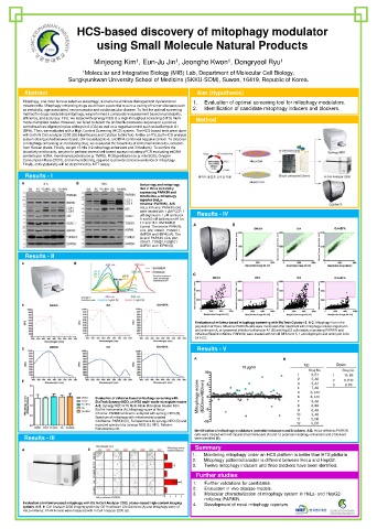
Identification of mitophagy modulators (potential enhancers and blockers). A-B. HeLa-mKeima::PARKIN
Results - III cells were treated with 640 natural small molecules (A) and 12 potential mitophagy enhancers and 3 blockers
were identified (B).
Summary
1. Monitoring mitophagy under an HCS platform is better than HTS platform
2. Mitophagy pattern/character is different between HeLa and HepG2.
3. Twelve mitophagy inducers and three blockers have been identified.
Further studies
1. Further validations for candidates.
2. Evaluation in vivo disease models.
3. Molecular characterization of mitophagy system in HeLa- and HepG2-
mKeima::PARKIN.
Evaluation of mKeima-based mitophagy with GE In Cell Analyzer 2200, a laser-based high-content imaging 4. Development of novel mitophagy reporters.
system. A-B. In Cell Analyzer 2200 imaging system by GE Healthcare Life Sciences (A) and Mitophagy score of
HeLa-mKeima::PARKIN cells were measured with In Cell Analyzer 2200 (B).