
Page 1 - F. Cell biology
P. 1
TMBIM6 (transmembrane BAX inhibitor motif containing 6) enhances autophagy
through regulation of lysosomal calcium
3
1
Hyun-Kyoung Kim , Geum-Hwa Lee , Kashi Raj Bhattarai , Myung-Shik Lee , Sung Hoon Back , Hyung-Ryong Kim , Han-Jung Chae 1
1
1
2
4
1 Department of Pharmacology and New Drug Development Research Institute, Chonbuk National University Medical School, Jeonju,
2 Severance Biomedical Science Institute and Department of Internal Medicine, Yonsei University College of Medicine, Seoul, School of
3
4
Biological Sciences, University of Ulsan, Ulsan, College of Dentistry, Dankook University, Cheonan, Republic of Korea
BACKGROUND AIM
TMBIM6 (transmembrane BAX inhibitor motif containing 6), a highly Recent findings indicated that TMBIM6 interacts with ITPR, which
conserved multi-transmembrane protein, has been identified as a may regulate steady-state [Ca ]ER, leading to the relatively low
2+
2+
suppressor of BAX-mediated cell death. TMBIM6 has been suggested mitochondrial calcium ([Ca ]mito) levels and reduced mitochondrial
to be a Ca 2+ channel-like protein that is integral to the intracellular bioenergetics, and ultimately autophagy. Independently, TMBIM6-
membranes of ER. The calcium-binding activity has been found to be specific regulation of a specific arm of ER stress involving
responsive to protons and other cations. The conserved aspartyl dyad ERN1/IRE-1α has also been reported in the context of secretory
(Asp171-Asp195) in an uncharacterized protein YetJ from Bacillus protein IgG and autophagy studies. Although there have been a
subtilis (BsYetJ) among TMBIM members regulates pH-dependent few studies on TMBIM6-associated autophagy regulation, the effect
calcium-binding and manages the channel pore opening and closing, of TMBIM6 on ER and lysosomal Ca 2+ signaling-associated
and Ca2+ translocation. The Ca permeating role of TMBIM6 lowers autophagy has not been studied yet. In the present study, we have
2+-
2+
the steady-state [Ca ]ER. investigated the role of TMBIM6 in lysosomal Ca 2+ signaling and
related autophagy.
METHODS
GCaMP3-ML-1 Ca 2+ imaging, Fura-2 Ca 2+ imaging, Oregon green 488 BAPTA-1 dextran (OG-BAPTA-dextran) or Rhod-dextran imaging,
Immunofluorescence assays, Immunohistochemistry, Proximity ligation assay (PLA), Real-time PCR analysis, and Autophagy flux detection were
performed.
RESULTS
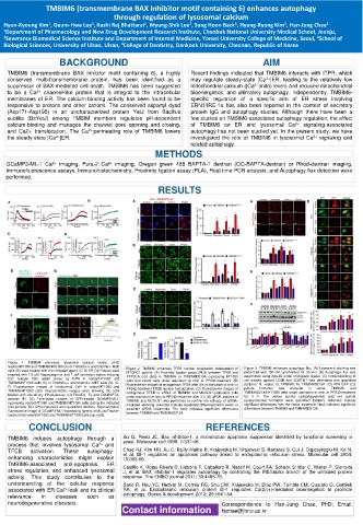
Figure 1. TMBIM6 enhances lysosomal calcium levels. (A-B)
Vector/HT1080 and TMBIM6/HT1080 (A) or Tmbim6+/+ and tmbim6-/- MEF Figure 2. TMBIM6 enhances TFEB nuclear localization independent of Figure 3. TMBIM6 enhances autophagy flux. (A) Lysosomal staining was
cells (B) were treated with the indicated agent. (C-D) ER Ca 2+ stores were MTORC1 activity. (A) Proximity ligation assay (PLA) between TFEB and performed with 100 nM LysoTracker for 30 min. (B) Autophagic flux was
emptied with 1-5 μM thapsigargin or and 1 μM ionomycin before inducing PPP3CA (red dots) in TMBIM6- or TMBIM6D213A expressing HT1080 determined using cyto-ID under microplate reader. (C) Immunoblotting of
Ca 2+ release from acidic stores by GPN in vector/HT1080 and cells and vector cells under starvation or torin or PP242-treatment. (B) cell lysates against LC3B and SQSTM1 was performed and quantified
TMBIM6/HT1080 cells (C) or Tmbim6+/+ and tmbim6-/- MEF cells (D). (E- Fluorescence images of endogenous TFEB after 3 h of starvation or torin or (bottom). N, vector; B, TMBIM6; M, TMBIM6D213A. (D) RFP-GFP-LC3
F) Fluorescence images of intraluminal Ca 2+ in vector/HT1080 and PP242-treatment TFEB nuclear translocation. (C) Fluorescence images of puncta formation was analyzed in vector, TMBIM6, and
TMBIM6/HT1080 cells. Representative images were showing the cells endogenous TFEB in siRNA of TMBIM6 and MCOLN1-pretreated cells TMBIM6D213A/HT1080 cells under starvation or torin or PP242-treatment
loaded with low-affinity Rhod-dextran (LA-RhodDx, E) and OG-BAPTA- under starvation or torin or PP242-treatment after 3 h. (D) qPCR analysis of for 3 h. The yellow puncta (autophagosome) and red puncta
dextran (F). (G) Time-lapse images of GPN-treated GCaMP3-ML1- TMBIM6 and MCOLN1 was performed to confirm the efficacy of siRNA- (autolysosome) formation were quantified (bottom). Asterisks indicate
expressing vector/HT1080 and TMBIM6/HT1080 cells during the indicated mediated silencing. Asterisks indicate significant differences from vector or significant differences from the vector treatment. Hash indicates significant
time periods. The GPN responses were quantified (bottom). Representative scramble siRNA treatments. The hash indicates significant differences differences between TMBIM6 and TMBIM6D213A.
fluorescence image of GCaMP3-ML1-expressing (green) and LysoTracker- between TMBIM6 and TMBIM6D213A.
loaded (red) vector/HT1080 and TMBIM6/HT1080 cells (up, right).
CONCLUSION REFERENCES
TMBIM6 induces autophagy through a Xu Q, Reed JC. Bax inhibitor-1, a mammalian apoptosis suppressor identified by functional screening in
process that involves lysosomal Ca 2+ and yeast. Molecular cell 1998; 1:337-46.
TFEB activation. These autophagy- Chae HJ, Kim HR, Xu C, Bailly-Maitre B, Krajewska M, Krajewski S, Banares S, Cui J, Digicaylioglu M, Ke N,
enhancing characteristics might explain et al. BI-1 regulates an apoptosis pathway linked to endoplasmic reticulum stress. Molecular cell 2004;
15:355-66.
TMBIM6-associated anti-apoptosis, ER Castillo K, Rojas-Rivera D, Lisbona F, Caballero B, Nassif M, Court FA, Schuck S, Ibar C, Walter P, Sierralta
stress regulation, and enhanced lysosomal J, et al. BAX inhibitor-1 regulates autophagy by controlling the IRE1alpha branch of the unfolded protein
activity. This study contributes to the response. The EMBO journal 2011; 30:4465-78.
understanding of the cellular response Sano R, Hou YC, Hedvat M, Correa RG, Shu CW, Krajewska M, Diaz PW, Tamble CM, Quarato G, Gottlieb
associated with ER Ca leak and its clinical RA, et al. Endoplasmic reticulum protein BI-1 regulates Ca(2)(+)-mediated bioenergetics to promote
2+
relevance in diseases such as autophagy. Genes & development 2012; 26:1041-54.
neurodegenerative diseases. Correspondence to Han-Jung Chae, PhD; Email:
Contact information hjchae@jbnu.ac.kr