
Page 29 - D. Cancer biology
P. 29
CD133-containing microvesicles transport SNAI1 and
promote cell motility in colon cancer
Jinsuk Pyo and Jesang Ko
Department of Life Sciences, Korea University Graduate School, Seoul 02841, South Korea
ABSTRACT
Extracellular vesicles (EV) derived from tumor microenvironment carry numerous bioactive molecules to deliver to recipient cells. Microvesicles (MV) are a type of EV that are released from the cell membrane and play an important role
in cell-to-cell communication. Epithelial-to-mesenchymal transition (EMT) is considered as a pivotal procedure of carcinogenesis, cancer cell mobility, and metastasis. However, it is not yet clear whether EMT-related molecules are
delivered by MVs. We reported that CD133 modulates MV release and oncoprotein trafficking in colon cancer. In this study, we investigated the role of CD133-containing MVs in the transport of EMT-related molecules in colon cancer.
We examined the components of MVs derived from CD133-positive and –knockdown HCT116 colon cancer cells. Among various EMT-related proteins, only SNAI1 was carried by MVs from CD133-positive cells and transported to
recipient cells. The delivered SNAI1 up-regulated the expression of ZEB1 and Vimentin in recipient cells. In addition, CD133-containing MVs promoted migration and invasion of recipient cells. However, MVs from CD133-knockdown
cells did not affect the motility of recipient cells. These results indicate that CD133-containing MVs are involved in regulating cell motility by delivering SNAI1.
BACKGROUND AIM
• Microvesicles (MV)
Purpose of the study is to understand the role of CD133-containing MVs in colon cancer
MVs are a type of secreted extracellular vesicles which are 100 – 1000nm in diameter (1). Various cargo molecules, including
cholesterol, protein, and RNA, are segregated during the MV formation (2). Furthermore, MVs deliver various signals by transporting • To understand the role of CD133-containing MVs in the transport of EMT-related
RNAs and proteins to target cells (3). Therefore, MVs are considered as a cell-to-cell communication tool, either short or long distance. molecules in colon cancer
• To understand the role of CD133-containing MVs in regulating cell motility of recipient
• SNAI1
cells
SNAI1 is a zinc finger protein associated with the EMT process. SNAI1 binds to the promoter region of Vimentin and ZEB1, and up- • To investigate whether MVs can be used as a tool for diagnosing and treating cancer
regulates their transcription (4). Along with Vimentin and ZEB1, SNAI1 down-regulates the E-cadherin transcription and promote the metastasis in colon cancer
EMT process (4,5).
RESULTS
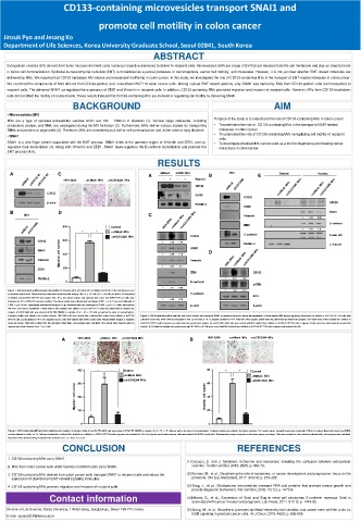
A C A shMock shCD133#4 B MVs E Cytosol Nucleus
HEK-293A +shMock MVs +shCD133#4 MVs - + - + Hypoxia
CD133
CD133
SNAI1 - -
CD133
β-actin CD133
Flotillin-2
SNAI1
B D SNAI1
MVs C Vimentin
1 1.23 0.58
β -catenin
800 Control ZEB1
1 1.75 1.6
*** *** +shMock MVs E-cadherin
+shCD133#4 MVs CD133 Lamin A/C
Migration cell number 400 1 2.69 1.13
CD133 600 1 5.15 1.04 Flotillin-2 α -tubulin
SNAI1 SNAI1 D
Vimentin Vimentin
ZEB1 200 1 3.77 3.1
ZEB1 CD133
Flotillin-2 1 5.21 1.02
0 N-cadherin
p-ERK
1 2.4 0.58
Figure 1. CD133-containing MVs promote the motility of recipient cells. (A) Cell lysates of shMock and shCD133 (#1 and #4) were used 1 1.43 0.78
to determine protein level. Protein level was determined by western blot analysis. (B) 2.4 ⅹ 10 7 cells (6.0 ⅹ 10 6 cells per well in 150 mm plates) E-cadherin ERK
of shMock and shCD133 HCT116 were plated. After 48 h, the culture medium was replaced with serum free RPMI 1640 and cells were
incubated for 24 h in 20% O2 normoxia condition. The culture media were collected and centrifuged at 500 ⅹ g for 5 min and additionally at 1 0.58 0.78
3,000 ⅹ g for 10 min. Supernatants were filtered through a 0.8 µm membrane filter and centrifuged at 20,000 ⅹ g for 1 h. Pellets were washed β-actin β-actin
with PBS and used for experiment. Protein level of MVs isolated from shMock or shCD133 HCT116 cells was determined by western blot
analysis. (C) HEK-293A cells were plated in 0.5% FBS DMEM at a density of 3.0 - 3.5 ⅹ 10 4 cells per well on the insert of transwell plates.
Complete medium was added to the bottom chamber. HEK-293A cells were treated with or without MVs isolated from shMock or shCD133 Figure 2. CD133-containing MVs obtained from colon cancer cells transport SNAI1 to recipient cells and induce the expression of downstream EMT-related signaling molecules. (A) shMock or shCD133 HCT116 cells were
HCT116 cells and incubated for 48 h. For migration assay, cells were stained with 0.05% crystal violet. Representative images of migration cultured in serum free RPMI 1640 and incubated in 20% O 2 normoxia or 1% O 2 hypoxia condition for 24 h. After MVs were isolated, protein level was determined by western blot analysis. (B) Protein level of MVs isolated from shMock or
assay are shown. Cells were counted from five randomly picked fields, and averages were calculated. All p values were obtained using an shCD133 HCT116 cells in hypoxia was determined by western blot analysis. (C and D) HEK-293A cells were treated with MVs isolated from shMock or shCD133 HCT116 cells in hypoxia. Protein level was determined by western blot
unpaired two-tailed Student’s t test. ***p < 0.001. analysis. (E) Cellular fractionation was performed using HEK-293A cell. Cells were treated with MVs isolated from shMock or shCD133 HCT116 cells in hypoxiaand incubated for 48h
A HEK-293A +shMock MVs +shCD133#4 MVs B HEK-293A +shMock MVs +shCD133#4 MVs
400 ** * Control 80 ** ** Control
+shMock MVs +shMock MVs
+shCD133#4 MVs +shCD133#4 MVs
300
Migration cell number 200 Invasion cell number 60 40
100
0 20 0
Figure 3. CD133-containing MVs promote migration and invasion of recipient cells. (A and B) HEK-293A cells were plated in 0.5% FBS DMEM at a density of 3.0 - 3.5 ⅹ 10 4 cells per well on the insert of transwell plates. Complete medium was added to the bottom chamber. For invasion assay, transwell inserts were coated with 100 µl of matrigel, diluted with serum free DMEM,
and then allowed to solidify for 4 h. Cell were treated with or without MVs isolated from shMock or shCD133 HCT116 cells in hypoxia and incubated for 48 h. For migration and invasion assays, cells were stained with 0.05% crystal violet. Representative image of migration and invasion assays are shown. Cells were counted from five randomly selected fields, and averages were calculated.
All p values were obtained using an unpaired two-student’s t test. * p < 0.05, ** p < 0.01.
CONCLUSION REFERENCES
1. CD133-containing MVs carry SNAI1.
(1) Cocucci, E. and J. Meldolesi, Ectosome and exosomes: shedding the confusion between extracellular
2. MVs from colon cancer cells under hypoxia conditions also carry SNAI1. vesicles. Trends Cell Biol, 2015. 25(6): p. 364-72.
3. CD133-containing MVs derived from colon cancer cells transport SNAI1 to recipient cells and induce the (2) Surman, M., et al., Deciphering the role of ectosomes in cancer development and progression: focus on the
expression of downstream EMT-related signaling molecules. proteome. Clin Exp Metastasis, 2017. 34(3-4): p. 273-289.
4. CD133-containing MVs promote migration and invasion of recipient cells. (3) Skog, J., et al., Glioblastoma microvesicles transport RNA and proteins that promote tumour growth and
provide diagnostic biomarkers. Nat Cell Biol, 2008. 10(12): p. 1470-6.
Contact information (4) Mikami, S., et al., Expression of Snail and Slug in renal cell carcinoma: E-cadherin repressor Snail is
associated with cancer invasion and prognosis. Lab Invest, 2011. 91(10): p. 1443-58.
Division of Life Science, Korea University, 1 Anam-dong, Sungbuk-gu, Seoul 136-701, Korea. (5) Wang, M., et al., N-cadherin promotes epithelial-mesenchymal transition and cancer stem cell-like tarits via
ErbB signaling in prostate cancer cells. Int J Oncol, 2016. 48(2): p. 595-606.
E-mail : pyojs9320@korea.ac.kr