
Page 117 - D. Cancer biology
P. 117
Activation of the Sympathetic Nerve System Weakens Anti-Tumoral Immunity
via Beta 2 Adrenergic Receptors in Osteoblast and Myeloid-Derived Suppressor Cells,
Contributing to Bone Metastasis Progression
1
1
Eun Jung Lee , Kyoung Jin Lee , Serk In Park 1
1 Department of Biochemistry and Molecular Biology, Korea University College of Medicine, Seoul, Korea
Background & Aim
Bone metastasis is the most significant factor affecting the patient survival of breast cancer patients, but no cure or preventive approaches are currently available. One factor that potentially affects
metastatic bone microenvironment is the sympathetic nerve system (SNS). The SNS regulates bone homeostasis via β2 adrenergic receptor (AR) on osteoblasts, and subsequently chronic stress-
induced SNS activation was shown to promote bone metastasis tumor growth, osteolysis, angiogenesis by osteoblastic cytokine expression in immunocompromised mouse models. On the other hand,
we previously demonstrated that osteoblast activation could lead to myeloid-derived suppressor cells (MDSC) activation, contributing to orthotopic prostate tumor growth. Therefore, we further
investigated the effects of SNS activation on immune cells via β2 AR on osteoblasts in the bone microenvironment using immune-competent mouse models of breast cancer bone metastasis.
Methods
A chronic immobilization stress (CIS) mouse model, one of the well-established models for the sympathetic nerve system activation in mice, was used. Mice were challenged with daily two-hour CIS
and tumor cells were implanted in the tibia on day-15. To block the SNS activation, a chemical selective inhibitor of β2 AR was used. At the endpoint analysis, tumors measured by bioluminescence
imaging. Tumor bearing tibia, tumor-naïve tibia or spleen were pulverized and performed flow cytometry. Additionally, to confirm the effects on MDSCs by β2 AR -stimulated osteoblast in vitro, we
cocultured both bone marrow cells and osteoblasts containing with or without β2 AR agonist, isoproterenol and then performed RT-qPCR on bone marrow cells.
Results
A. B. 1 A. B. C. 3
CIS ± ICI or Vehicle Tx (25 d.) P>0.05 P>0.05
P<0.05 P>0.05 P>0.05
P<0.05 CIS ± ICI or Vehicle 30 95
Bioluminescence
Balb/c mice 14 d. 11 d. 30
(Tumor Size) 25 Balb/c 14 d. Bioluminescence & 20 90
15
15
4T1 intra-tibial 20 mice Flow Cytometry ×10 4 Photon/S 25 % CD45 + cells 85
tumor inj. ×10 8 Photon/S 10 4T1 intra-tibial 10 80
Chronic Con 5 tumor inj. 5 75
Immobilization 0 Con Veh ICI 70 Con Veh ICI
Stress (CIS) 0
2 hours daily Veh Con Veh ICI CIS CIS
CIS CIS
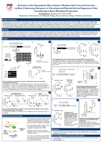
ICI CIS Treatment after tumor inoculation did not increase MDSC nor tumor growth.
*ICI 118,551 (A) Experimental scheme. 4T1 cancer cells were inoculated in the tibia followed by CIS in
1x10 6 5x10 7 A selective β2 AR antagonist combination with ICI118,551 for two weeks. (B) Quantification of the BLI signal intensity at the
[photon/sec] endpoint. (C) Flow cytometric quantification of CD45 + CD11b + Gr-1 + MDSCs, in the tumor bearing
bone marrow.
C. D.
4T1 Cancer Cell
Proliferation in vitro 4
A. * Positive Control: Tumor Implantation C. P<0.0001
ICI 118,551 (5 μM)
Adrb1 10 8 P˃0.05 Vehicle Control or CIS 80 P<0.0001
Adrb2 Cell Count (Fold) 6 Balb/c Flow Cytometry & 60
Adrb3 4 mice 7 d. +7 d. T cell assay CFSE-labeled T cells % 40
GAPDH 2 20
0
0 1 2 days B. 0
CD11b + Gr-1 + cells CD11b+Gr-1+ Naïve - 1 - - -
4T1 tumor growth was promoted by chronic immobilization stress and reversed by EdU+ Cells T/ Gr-1 high CIS (2wk) - - 1 - -
osteoblastic β2 AR inhibition. P<0.01 P<0.01 cells ratio - - 1 0.5
(A) Experimental scheme. Chronic immobilization stress (CIS) in combination with ICI118,551 100 40 P.C -
were treated for 14 days and 4T1 cancer cells were implanted in the proximal tibiae, followed by P<0.01 P<0.01
tumor growth for 11 days. (B) Quantification of the BLI signal intensity (C) A semi-quantitative 80 30 CIS treatment increased proliferation and
RT-PCR analysis of β-ARs in murine pre-osteoblast cells and two murine breast cancer cells. (D) 60 function of MDSC in the bone marrow
4T1 cancer cell proliferation assay for 2 days in the presence of β2-AR antagonist. % CD45 + cells 40 % CD45 + cells 20 (A) Experimental scheme. Female Balb/c mice
were treated with CIS for 7-14 days. (B) Flow
2 20 10 cytometric quantification of CD45 + CD11b + Gr-1 +
MDSCs or EDU + MDSCs in the bone marrow.
Tumor-Bearing Bone Spleen 0 0 (C) Flow cytometric quantification of CFSE-
Con 1 2 P.C Con 1 2 P.C
CD11b + Gr-1 + cells Gating: CD45 + Viability + CD11b + Gr-1 + cells Gating: CD45 + Viability + CIS (wk) CIS (wk) labelled CD3 + T cells after 3 days co-cultured
P<0.01 P>0.05 with Gr-1 high+ MDSCs.
100 Con CIS 10 8 Con CIS 5 6
95
% CD45 + cells 90 Gr-1 % CD45 + cells 6 4 2 Gr-1 A. CIS ± ICI or Vehicle Arg1 P<0.01
85
P<0.01
80
75
Con CIS CD11b 0 Con CIS CD11b C57BL/6 10 d. Flow Cytometry 40 P<0.05
mice
20
CIS-induced Bone Metastases Contained Increased CD11b + Gr1 + MDSC in tumor-bearing Relative mRNA level 60 5 4
bone microenvironment, not in spleen. B. CD11b + Gr-1 + cells 3 2
Flow cytometric quantification of CD45 + CD11b + Gr-1 + MDSCs in the tumor bearing bone marrow 1
and spleen. P<0.01 A selective of β2 GM-CSF 0 - + + + +
adrenergic receptor (AR)
60 P<0.05 antagonist reversed CIS- Adrb2 agonist - - + - +
Summary and Conclusions 55 induced MDSCs increase. Osteoblast - - - + +
• The sympathetic nerve system (SNS) activation % CD45 + cells 50 (A) Experimental scheme. Arginase immunosuppressive function
C57BL/6 mice were treated
45
(via chronic immobilization stress) increases 40 with CIS±ICI for 10 days. was increased by β2 AR-Stimulated
1) MDSC expansion and immuno-suppressive 35 (B) Flow cytometric Osteoblasts.
function; and 2) bone metastasis tumor growth. 30 quantification of Relative Arg1 mRNA level on bone
Con Veh ICI CD45 + CD11b + Gr-1 + MDSCs marrow cells co-cultured with osteoblasts
• The pro-tumorigenic function of SNS activation CIS in the bone marrow. in presence of 20 ng/mL of GM-CSF and
is mediated by β2 adrenergic receptors 10 uM of isoproterenol or not.
expressed in osteoblasts and/or MDSC.
Reference
• Candidates derived from β2 AR-activating
osteoblast will be found and confirmed its effect • • Leptin regulation of bone resorption by the sympathetic nervous system and CART. Elefteriou F et al. 2005 Nature.
Stimulation of Host Bone Marrow Stromal Cells by Sympathetic Nerves Promotes Breast Cancer Bone Metastasis in Mice.
on MDSC regulation. Campbell JP et al. Plos Biol 2012
• In conclusion, hyperactivity of the sympathetic Acknowledgements & Contact information
nerve system (e.g. via mental stress) tips the
anti-tumoral immunity balance in bone towards • Financial Supports: the National R&D Program for Cancer Control, the Ministry of Health and Welfare, the Republic of
bone metastasis progression. • Korea (HA17C0040).
Email: ejlee3365@korea.ac.kr