
Page 115 - D. Cancer biology
P. 115
Pristimerin isolated from the Celastraceae and Hippocrateaceae families is a naturally occurring quinone methide triterpenoid. Our previous indicates the potential value
of pristimerin in suppressing colon tumorigenesis via regulating the AKT/FOXO3a signaling pathway. MicroRNAs (miRNAs ) are small, non-protein-coding RNAs that can
function as tumor suppressors or oncogenes. Deregulation of miRNA expression has been reported in various cancer including ovarian cancer. However, modulation of
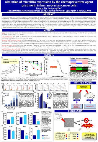
miRNA expression by chemopreventive phytochemicals remains to be defined. In the present study, we investigated whether prisitmerin treatment altered miRNA levels
in SKOV-3 cells to clarify the detailed mechanism of the anticancer effect of pristimerin. Pristimerin caused a significant increase of miR-195-5p and miR-374b-5p
expression in SKOV3 cells, while significantly impeding cell proliferation and stimulating apoptosis. In addition, pristimerin treatment reduced the expression PSAT1 and
BIRC3 gene, which are representative targets of miR-195-5p and miR-374b-5p, respectively. Since PSAT1 and BIRC3 has oncogenic activity, our results indicated that
induced expression of miR-195-5p and miR-374b-5p by pristimerin treatment.
Cells: Human ovarian cancer cells, SKOV-3 cells obtained from the Korean Cell Bank. The cells were cultured in RPMI-1640 containing 5% FBS, 100 U/ml penicillin, and
100 μg/ml streptomycin at 37℃ in 5% CO 2 .
Cell viability assay: The effects of pristimerin on cell viability were tested using the CCK-8 Assay, which used colorimetry to count the number of viable cells. The
absorbance was read at 450 nm in a microplate reader.
Apoptosis and Cell cycle analysis: SKOV-3 cells were seeded at a density of 3×10 cells. Cells were treated with indicated concentration of pristimerin for 24 hours.
6
Apoptotic cells were detected using a Muse Annexin V and Dead Cell kit and Muse Cell analyzer. Pristimrin treated cells were collected and fixed in ice-cold 70% ethanol
and stored at 4℃ overnight. Cell cycle status was determined by measuring cellular DNA content after staining with Muse cell cycle kit and Muse Cell analyzer.
6
MiRNA and mRNA array: SKOV-3 cells were seeded at a density of 1×10 cells and treated with pristimerin IC 50 for 24 hours. Total RNAs were extracted from cells using
TRI solution(BIOSCIENCE TECHNOLOGY) and commissioned by the Bio-core company for the miRNA and gene arrays.
MiRNA RCR and Gene qRT-PCR: MiRNAs were extracted from cells using mirVana™ miRNA Isolation Kit, with phenol (Invitrogen) following manufacturer’s instructions.
MiRNA expression was analyzed by real-time PCR using TaqMan™ Universal PCR Master Mix, no AmpErase™ UNG. mRNAs were extracted from cells using TRI
solution(BIOSCIENCE TECHNOLOGY) and gene expression was analyzed by real-time qPCR using Go Taq qPCR Master Mix kit.
μ
μ μ μ μ μ μ μ μ μ μ
μ
μ
μ
μ
μ μ
μ