


Page 35 - ebook
P. 35
Choline kinase alpha 2 acts as a protein kinase
to promote lipolysis of lipid droplets
Su Hwan Park and Jong-Ho Lee
Department of Health Sciences, The Graduate School of Dong-A University, Busan, 49315, Republic of Korea
ABSTRACT INTRODUCTION CONCLUSIONS
Lipid droplets are important for cancer cell growth and 1. Lipid droplets are involved in many aspects of cellular activities and are
survival. However, the mechanism underlying the associated with cancer cell survival (1).
initiation of lipid droplet lipolysis is not well understood. 2. Cells use these stored lipids as needed for a variety of functions,
We demonstrate here that glucose deprivation induces
the binding of choline kinase (CHK) α2 to lipid including energy production via fatty acid oxidation (also known as β-
droplets, which is sequentially mediated by AMPK- oxidation) (2).
dependent CHKα2 S279 phosphorylation and KAT5- 4. The heat shock cognate protein of 70 kDa (Hsc70) binds to PLIN2 and
dependent CHKα2 K247 acetylation. Importantly, PLIN3 to induce their lysosomal degradation, which facilitates lipid droplet
CHKα2 with altered catalytic domain conformation association with cytosolic lipase ATGL and macroautophagy (3, 4).
functions as a protein kinase and phosphorylates
PLIN2 at Y232 and PLIN3 at Y251. The However, how lipolysis of lipid droplets is initiated is relatively unknown.
phosphorylated PLIN2/3 dissociate from lipid droplets 5. CHK is expressed in at least three isoforms; CHKα1, CHKα2, and CHKβ
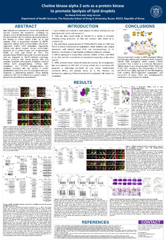
and are degraded by Hsc70-mediated autophagy, (5, 6). CHK can form homodimers or heterodimers depending on the tissue Glucose deprivation results in AMPK-mediated CHKα2
thereby promoting lipid droplet lipolysis, fatty acid type (7). S279 phosphorylation and subsequent KAT5-mediated
oxidation, and brain tumor growth. In addition, levels of CHKα2 K247 acetylation, which induces CHKα2
CHKα2 S279 phosphorylation, CHKα2 K247 6. CHKα promotes tumor cell proliferation and survival. Its overexpression conversion from a dimer to a monomer. Monomeric
acetylation, and PLIN2/3 phosphorylation are has been detected in 40%–60% of human tumors and is correlated with CHKα2 binds to and phosphorylates PLIN2 Y232 and
positively correlated with one another in human prognosis in early-stage non-small cell lung cancer, hepatocellular PLIN3 Y251, leading to the disassociation of PLIN2
glioblastoma specimens and are associated with poor carcinoma (HCC), and prostate cancer (8, 9, 10). However, the and PLIN3 from lipids, exposure of the CMA-targeting
prognosis in glioblastoma patients. These findings mechanisms underlying CHKα-enhanced tumor progression still need to be motif enabling Hsc70-dependent degradation, and
underscore the role of CHKα2 as a protein kinase in elucidated. recruitment of lipase ATGL and autophagosome
lipolysis and glioblastoma development. machinery to lipid droplets for lipolysis.
RESULTS
Figure 3. Monomeric CHKα2 binds to and
phosphorylates PLIN2/3
(A) Huh7 cells expressing the indicated CHK proteins
with or without expression of KAT5 shRNA-1 were
stimulated by glucose deprivation for 1 h. A streptavidin
pull-down assay was performed. WCL, whole-cell lysate.
(B and D) Huh7 cells expressing the indicated HA- or
FLAG-tagged CHKα2 proteins were stimulated by
glucose deprivation for 1 h. Immunoprecipitation
analyses (B) or a streptavidin pull-down assay (D) was
performed. (C) The interaction of E97 and K247 between
the monomers of a CHKα2 dimer is shown. (E) Huh7
cells were stimulated with glucose deprivation for 1 h.
Immunoprecipitation with indicated antibodies were
performed. (F and H) Huh7 cells expressing FLAG-
PLIN2, FLAG-PLIN3, WT His-CHKα2, or the indicated
CHKα2 mutants were stimulated by glucose deprivation
for 1 h. Immunoprecipitation with anti-FLAG M2 agarose
beads and a Ni-NTA agarose bead pull-down assay were
performed. (G) Two CHKα2 molecules in dimeric form.
The sequence of the interface is shown. The
hydrophobic and hydrophilic residues are labeled in blue
and purple, respectively. The dimer interface is enlarged.
The side chain of I186 and L187 is shown in yellow. (I)
An in vitro kinase assay was performed by mixing
bacterially purified active AMPK (AMPKα1/β1/γ1) with
the indicated purified FLAG-CHKα2 proteins. The CHKα2
Figure 1. CHKα2 binds to lipid droplets and is required for lipid droplet lipolysis proteins were then pulled down, washed, and incubated
(A) The indicated cells were stimulated by glucose deprivation for 1 h. Expression of with bacterially purified His-KAT5 for an in vitro acetylation assay. The CHKα2 proteins were then pulled down, washed, and eluted with FLAG peptide. Bacterially purified WT His-PLIN2 or WT His-
CHKα in the whole-cell lysate (WCL), cytosol (Cyto), and lipid droplet (LD) fractions was PLIN3 protein, which were immobilized on Ni-NTA beads, were then mixed with CHKα2 proteins for an in vitro kinase assay in presence of [γ- P]ATP. Autoradiography and immunoblotting analyses
32
examined. Samples were normalized to cell number. (B) Huh7 cells were stimulated by were performed. (J) The AMPK-phosphorylated and KAT5-acetylated WT FLAG-CHKα2 protein, described in (I), was mixed with the immobilized, purified, and the indicated His-PLIN2 or His-PLIN3
glucose deprivation for 1 h and stained with BODIPY, DAPI, or antibodies recognizing proteins in the presence of [γ- P]ATP for in vitro kinase assay. Autoradiography and immunoblot analyses were performed. (K) Bacterially purified WT His-PLIN2 or WT His-PLIN3 protein, which was
32
CHKα or CHKβ. Boxed areas are enlarged and placed underneath. The relative intensity immobilized on Ni-NTA beads, was mixed with bacterially purified WT FLAG-CHKα2, acetylation-mimicking mutant CHKα2 K247Q, or kinase-dead acetylation-mimicking mutant CHKα2
of green (BODIPY) or red (CHKα or CHKβ) fluorescent signals along the white oblique K247Q/D330A/E332A in the presence of ATP for an in vitro kinase assay. (L) Bacterially purified WT His-PLIN2, WT His-PLIN3 or the indicated mutant proteins, which was immobilized on Ni-NTA
line were measured using FV10 ASW (version 3.1). Arrows indicate the colocalized beads, was mixed with bacterially purified FLAG-CHKα2 K247Q protein in the presence of ATP for an in vitro kinase assay. (M) Huh7 cells expressing the indicated His-PLIN2 or His-PLIN3 proteins
green and red signal peaks. Scale bars, 15 μm (original images) and 3 μm (enlarged were stimulated by glucose deprivation for 1 h. A Ni-NTA agarose bead pull-down assay was performed. (N) His-PLIN2 and FLAG-PLIN3 were exogenously expressed in Huh7 or U87 cells that were
images). (C) Huh7 cells, with or without expression of CHKα2 shRNA-1 or CHKβ with expression of CHKα2 shRNA-1 and the indicated rCHKα2 proteins. These cells were stimulated by glucose deprivation for 1 h. Immunoprecipitation with anti-FLAG M2 agarose beads and a Ni-
shRNA, were stimulated by glucose deprivation for 1 h. Expression of the indicated NTA agarose bead pull-down assay were performed.
proteins was examined in purified lipid droplet or WCL samples. Lipid droplet samples
were normalized to triglyceride level. (D) Huh7 cells, with or without expression of
CHKα2 shRNA-1 or CHKβ shRNA, were stimulated by glucose deprivation for 1 h. The Figure 4. CHKα2-mediated PLIN2/3 phosphorylation leads to
cells were stained with BODIPY, DAPI, or antibodies recognizing Beclin1 or ATGL. the recruitment of Hsc70, ATGL, and autophagosome
Boxed areas are enlarged and placed underneath. Scale bars, 10 μm (original images) machinery to lipid droplets
and 3 μm (enlarged images). (A and B) Huh7 cells with expression of FLAG-PLIN2, His-PLIN3,
CHKα2 shRNA-1, and reconstituted expression of the indicated
rCHKα2 proteins were stimulated by glucose deprivation for 1 h.
Immunoprecipitation with anti-FLAG M2 agarose beads and a Ni-
NTA agarose bead pull-down assay was performed. WCL, whole-
cell lysate. (C) Huh7 cells expressing HA-Hsc70, WT FLAG-PLIN2,
WT FLAG-PLIN3, or their indicated mutants were stimulated by
glucose deprivation for 1 h. Immunoprecipitation with anti-FLAG M2
agarose beads was performed. (D) U87 cells and U87 cells with
knockin expression of CHKα2 S279A, CHKα2 K247R, or PLIN2/3
Mut were stimulated by glucose deprivation for 2 h. Cell were
stained with BODIPY, DAPI, and antibodies recognizing PLIN2
(upper panel) and PLIN3 (lower panel). Boxed areas are enlarged
and placed underneath. Scale bars, 10 μm (original images) and
2.5 μm (enlarged images).
Figure 5. CHKα2-mediated lipo-lysis of lipid droplets promotes tumor cell survival Figure 6. CHKα2-mediated lipolysis of lipid droplets promotes brain tumor growth
(A) U87 cells and U87 cells with knockin expression of CHKα2 S279A, CHKα2 K247R, or (A) U87 cells and U87 cells with knockin expression of CHKα2 S279A, CHKα2 K247R, PLIN2/3
PLIN2/3 Mut were incubated with or without glucose-free DMEM medium for 2 h. The levels of Mut, or expression of ATGL or Beclin1 shRNA were intracranially injected into athymic nude mice.
cellular glycerol, cellular fatty acid, acetyl-CoA, and ATP were examined. Results were Two weeks after tumor cell injection, 0.2 mL of 2-DG (500 mg/kg) was intraperitoneally injected
Figure 2. AMPK- and KAT5-mediated modification of CHKα2 promotes the binding
of CHKα2 to lipid droplet normalized to the number of cells. (B) U87 cells and U87 cells with knockin expression of CHKα2 daily for 14 days (n = 7). The mice were euthanized, and tumor growth was measured.
(A) Huh7 cells were stimulated with glucose deprivation for 1 h. (B) In vitro kinase S279A, CHKα2 K247R, or PLIN2/3 Mut were incubated with BSA-conjugated 13C18-oleic acid Hematoxylin and eosin-stained coronal brain sections show representative tumor xenografts. (B)
assays were performed by mixing purified bacterially expressed active His-AMPK (0.3 mM) for 12 h, then stimulated by glucose deprivation for 2 h. The levels of 13C-acetyl-CoA, Mouse tumor tissues were subjected to frozen sectioning. The slides were stained using oil red O
(AMPKα1/β1/γ1) with WT GST-CHKα2, GST-CHKα2 S279A, or GST-CHKβ in the 13C-citrate, and 13C-succinate were measured. The results were normalized to the number of (n = 7). (C) Sixty GBM samples with survival time information were included and categorized into
presence of [γ- P]ATP. Autoradiography and immunoblot analyses were performed. (C) cells. (C) U87 cells and U87 cells with knockin expression of CHKα2 S279A, CHKα2 K247R, or high (top 50%, n = 30) or low (bottom 50%, n = 30) IHC score groups on the basis of the IHC
32
Huh7 cells were treated with 5 μM compound C for 30 min and stimulated with glucose PLIN2/3 Mut were incubated with 25 mM 2-DG for 48 h. (D) U87 cells and U87 cells with knockin scores of PLIN2 pY232, PLIN3 pY251, CHKα2 pS279, or CHKα2 AcK247. Kaplan-Meier plots of
deprivation for 1 h or treated with 0.5 mM A769662 for 30 min. LD, lipid droplets. (D) expression of CHKα2 S279A, CHKα2 K247R, or PLIN2/3 Mut were stimulated by glucose the overall survival time were performed and compared. p value was calculated using the log
Huh7 and U87 cells expressing WT FLAG-CHKα2 or FLAG-CHKα2 S279A were deprivation for 8 h. Cell death was measured by TUNEL assay. ∗∗p < 0.01. ∗p < 0.05, ∗∗p < rank test.
stimulated by glucose deprivation for 1 h. Immunoprecipitation of whole-cell lysates with 0.01, and ∗∗∗p < 0.001.
anti-FLAG M2 agarose beads was performed. (E) Huh7 cells were stimulated with
glucose deprivation for 1 h. Immunoprecipitation with an anti-KAT5 antibody was
performed. (F) An in vitro kinase assay was performed by mixing purified AMPK REFERENCES CONTACT
(AMPKα1/β1/γ1) with bacterially purified WT GST-CHKα2 or GST-CHKα2 S279A
protein. GST-CHKα2 proteins were pulled down, washed with PBS, and incubated with Lab. Cancer metabolism, S11-0319, Natural
His-KAT5 protein. (G) An in vitro kinase assay was performed by mixing purified AMPK 1. S. Koizume, Y. Miyagi. Lipid droplets: a key cellular organelle associated with cancer cell survival under normoxia and hypoxia. Int. J. Mol. Sci., 17 (2016), p. 1430 Science Bldg. Dong-A University, 37, Nakdong-
2. A.D. Barbosa, D.B. Savage, S. Siniossoglou. Lipid droplet-organelle interactions: emerging roles in lipid metabolism. Curr. Opin. Cell Biol., 35 (2015), pp. 91-97
protein (AMPKα1/β1/γ1) with bacterially purified WT GST-CHKα2 or GST-CHKα2 K247R 3. S. Kaushik, A.M. Cuervo. Degradation of lipid droplet-associated proteins by chaperone-mediated autophagy facilitates lipolysis. Nat. Cell Biol., 17 (2015), pp. 759-770 Daero 550beon-gil saha-gu, Busan, Korea
proteins. GST-CHKα2 proteins were pulled down, washed with PBS, and incubated with 4. S. Kaushik, A.M. Cuervo. AMPK-dependent phosphorylation of lipid droplet protein PLIN2 triggers its degradation by CMA. Autophagy, 12 (2016), pp. 432-438 Tel: +82 51 200 6520
His-KAT5 for an in vitro acetylation assay. (H) Huh7 cells expressing KAT5 shRNA-1 5. E. Malito, N. Sekulic, W.C. Too, M. Konrad, A. Lavie. Elucidation of human choline kinase crystal structures in complex with the products ADP or phosphocholine. J. Mol. Biol., 364 (2006), pp. 136-151
6. X. Chen, H. Qiu, C. Wang, Y. Yuan, J. Tickner, J. Xu, J. Zou. Molecular structure and differential function of choline kinases CHKα and CHKβ in musculoskeletal system and cancer. Cytokine Growth
were stimulated by glucose deprivation for 1 h. (I) The indicated cells expressing WT Factor Rev., 33 (2017), pp. 65-72 Su Hwan Park
FLAG-CHKα2 or FLAG-CHKα2 K247R were stimulated by glucose deprivation for 1 h. 7. H. Liao, C. Aoyama, K. Ishidate, H. Teraoka. Deletion and alanine mutation analyses for the formation of active homo- or hetero-dimer complexes of mouse choline kinase-alpha and -beta. Biochim. E-mail: otter_sh96@naver.com
Immunoprecipitation with anti-FLAG M2 agarose beads was performed. Biophys. Acta, 1761 (2006), pp. 111-120 Jong-Ho Lee
8. A. Challapalli, et al. Exploiting altered patterns of choline kinase-alpha expression on human prostate tissue to prognosticate prostate cancer. J. Clin. Pathol., 68 (2015), pp. 703-709
9. S.A. Kwee, B. Hernandez, O. Chan, L. Wong. Choline kinase alpha and hexokinase-2 protein expression in hepatocellular carcinoma: association with survival. PLoS ONE, 7 (2012), p. e46591 E-mail: Topljh19@dau.ac.kr
10. A. Ramírez de Molina, et al. Expression of choline kinase alpha to predict outcome in patients with early-stage non-small-cell lung cancer: a retrospective study. Lancet Oncol., 8 (2007), pp. 889-897

