


Page 225 - ebook
P. 225
Isolation of lysosomal activator regulating lysosomal
damages and cell death
Heemin Park , Shin-Hyeon Ryu , Seo-hyun Kim , and Yong-Keun Jung *,1
1
1
1
1 Apoptosis and Molecular Disease Laboratory; School of biological science, Seoul National University, Seoul 08826, Korea
Abstract
Lysosomes contribute to maintaining cellular homeostasis via degradation and recycling of subcellular substances called autophagy, along with a protective action against foreign materials, such
as pathogens, and cellular protein aggregates. Therefore, cells avoid cytotoxicity of the invading pathogen or suppress cell death by degrading cytotoxic proteins and its aggregates through the
lysosomal function. We aimed to isolate small molecules that affect lysosomal damages and activities. In this study, we established a cell-based lysosomal activity assay and screened a bioactive
compound library to isolate new mediators affecting lysosomal damage. We generated HeLa/GFP-Galectin3 stable cell line and observed GFP-Gal3 puncta formation when exposed to
lysosomotropic LLOME. We isolated several compounds that affected the LLOME-induced GFP-Gal3 puncta formation. Among them, we found that mA receptor antagonist (mARA) rescued the
processing of cathepsin D and the lysosomal activity as assayed with DQ-red BSA. In addition, we found that mARA alleviated LLOME-induced apoptosis. Thus, we intend to broaden our
understanding on the lysosomal regulation to lysosomal disease models.
Result 3. mA receptor antagonists increase lysosomal activity
1. Establishment of a cell-based assay for identifying mediators
affecting regulation of intracellular lysosomal damage and activity
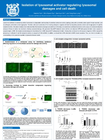
(A) HeLa cells were co-treated with 10 μM of
indicated compound and 10 μg/ml of DQ-
red-BSA. for 2 h. Then, 200 μM of LLoMe
was additively treated for 6 h. Images were
captured by fluorescence microscopy.
(B) Quantification of red fluorescence
intensity corrected by area in (A). Bars
represent mean ± S.E.M., compared with
nontreat(DMSO) control, one-way ANOVA.
n=3, cell number = 164-173
(C) Receptor M antagonist T03G06 dose
dependently increases lysosomal activity. 10
ug/ml of DQ-red-BSA was co-treated with
DMSO or 1/5/10 μM of compound T03G06 for
2 h, and then 200 μM of LLoMe was
additively treated for 6 h. Images were
captured by fluorescence microscopy.
(A) Schematic representation of the establishment of GFP-Gal3 expressing HeLa cell line and the LLoMe-
induced lysophagy and GFP-Gal3 puncta formation model. (B) Representative image of LLoMe-inudced GFP-
Gal3 puncta formation. HeLa/GFP-Gal3 cells were treated with 250 μM of LLoMe for 4 h. The images were 4. mA receptor antagonist T03G06(mARA) increases lysosomal activity
captured under fluorescence microscopy. (C) HeLa/GFP-Gal3 cells were plated in 12-well culture plate and
incubeated for 24 h. Cells were treated with or without 250 uM of LLoMe for 6 h. Cells were lysed with RIPA
buffer and whole cell lysate was subjected to western blot analysis using anti-Cathepsin D, anti-LC3, anti-GFP,
anti-β actin antibodies.
2. Screening strategy to isolate bioactive compounds regulating
lysosomal damage and activity
(A) HeLa cells were plated in 12-well culture plate. Cells were treated with indicated concentration of AQ-RA
741. After 2 h, 0, or 500 μM of LLoMe was added. After 6 h, cells were harvested and lysed with RIPA lysis buffer.
Samples were subjected to western blot analysis. (B,C) Quantification of western blot analysis. Relative band
intensity of conversion of LC3-I to LC3-II(B), and process of pro-Cathepsin D to mature-Cathepsin D was
measured.
5. T03G06 decreases LLoMe- 6. T03G06 decreased oAβ-
induced cell death induced cell death in HT22
C e ll d ea th as s a y (P I s ta in in g ) 1 6h
A -in d u c e d c y to to x ic ity (c e ll d e a th )
*** *
** *
2 0 n .s
*** * 2 5
)
(A) Schematic representation of the screening strategy to isolate bioactive compounds regulating lysosomal t (% 1 5 ) (% 2 0
damage and activity using Tocriscreen mini bioactive compound library. The compound library was prepared s h c M A
at 1 mM. HeLa/GFP-Gal3 cells were plated in 96-well culture plate and incubated for 24 h. Cells were treated e o 1 0 in e 1 5
with 10 μM each of the compounds for 2 h. Then, 200 μM of LLoMe was added. After 4 h, lysosomal damage H I+ lc a 1 0
was assessed based on GFP-Gal3 puncta formation. The compounds showing decreased number of GFP-Gal3 I/P 5 C I+
puncta were isolated. (B) Pharmacological feature of Tocriscreen 2.0 Mini compound library. Various type of P I/P P 5
target classes(left) and its primary actions(right) are shown. 0
S O S O 1 M 5 M 0
M D M 1 0 u M S O S O ) ) ) P F
Discussion C trl_ D T 03G 06 C trl_ D M D M 0 6 (1 M 0 6 (5 M 0 6 (1 0 M
LLO M e (200 M) T 0 3 G T 0 3 G T 0 3 G
⚫ Novel regulators of lysosomal activity were isolated via functional screening on HeLa/GFP-Gal3 cells were plated in 96-well o A (5 M )
Bioactive compound library culture plate and incubated for 24h. 1% DMSO HT22 cells were plated in 96-well culture plate
⚫ As putative activator of lysosome, T03G06 upregulates lysosomal activity and recovers or indicated concentration of AQ-RA 741 was with full media(10 % FBS). Indicated
lysosomal damage done by lysosomotropic agent, LLOMe. treated for 2 h, and then 0 or 200 uM of LLoMe concentration of compound and 5 μM of
oligomeric Aβ were co-treated with serum-free
⚫ TFEB phosphorylation at Ser211, which is the target residue of mTOR, was decreased was treated. After 16h, cells were stained with DMEM, and then cells were incubated for 48 h.
PI/Hoechst and cell death rate (PI/PI+Hoechst,
upon T03G06. This indicates that T03G06 may upregulate lysosomal activity through cell number) was calculated. Images were Cells were stained with PI+Cacein AM and cell
control of TFEB activity. captured by fluorescence microscopy. death rate was counted.

