


Page 169 - ebook
P. 169
Differentiation of Various Gastric Cancer Cell Lines
using Glycome Signatures by LC-MS/MS
1,2
1,2
3
1,2
Jihyeon Nam , Nari Seo , J Eugene Lee , and Hyun Joo An *
1. Asia-Pacific Glycomics Reference Site, Chungnam National University, Daejeon, Korea
2. Graduate School of Analytical Science and Technology, Chungnam National University, Daejeon, Korea
3. Center for Bioanalysis , Korea Research Institute of Standards and Science , Daejeon, Korea.
Overview Result
Gastric cancer (GC), the fifth most common cancer in the world, is the third most
lethal cancer in mortality. Cell lines are valuable tools for developing treatments Compositional Glycan Profiling by nano-LC/MS
that can minimize disparities in gastric cancer. Despite the high incidence of SNU-1 AGS
C/H-F&S
gastric cancer in Northeast Asia, cell lines derived from U.S.A. are the most
commonly used models for studying cancer biology, validating cancer targets and
C/H-S
for defining drug efficacy. In this study, glycan analysis from GC cell lines was
SNU-16 KATO-Ⅲ
performed to improve insight into gastric cancer. A total of 8 GC cell lines were Glycan Type C/H-F
selected, including 6 GC cell lines derived from Korea and Japan, which are
countries with a high incidence of gastric cancer. Glycom signatures of GC cell C/H
SNU-5 MKN-45
lines can serve as a valuable resource for developing targeted therapeutics in
HM
terms of ethnic and gender.
0 20 40 60
The need for cell lines from diverse ethnic backgrounds NCI-N87 MKN-74 NAPI (%)
Class
Statistical Information of Gastric cancer Gastric Cancer Cell Lines
1 SNU-1 5 AGS
Top 5 countries with gastric cancer incidence
New cases % 2 SNU-16 6 KATO-3
Rank Cancer diagnosed Age-standardised
of all cancers Rank Country 3 SNU-5 7 MKN-45
in 2018 rate per 100,000
AGS cell line ■ Core Type ■ High ■ Complex/Hybrid ■ C/H ■ C/H ■ C/H Sia & Fuc 4 NCI-N87 8 MKN-74
1 Lung 2,093,876 12.3% 1 South Korea 39.6 Mannose Type Fucosylation Sialylation
2 Breast 2,088,849 12.3% 2 Mongolia 33.1
3 Colorectal 1,800,977 10.6% 3 Japan 27.5
4 Prostate 1,276,106 7.5% 4 China 20.7 Different Expression Level of Glycans Distribution
5 Stomach 1,033,701 6.1% 5 Bhutan 19.4
6 Liver 841,080 5.0% 6 Kyrgyzstan 18.6
7 Oesophagus 572,034 3.4% 7 Chile 17.8 Heatmap Analysis Glycosylation Map
8 Cervix uteri 569,947 3.3%
8 Belarus 16.5
9 Thyroid 567,233 3.3%
9 Peru 16.1
*World Cancer Research Fund International As a common GC cell line, AGS has been
10 Bladder 549,393 3.2%
10 Vietnam 15.9
used widely in GC-related studies
Aberrant glycosylation as one of the hallmarks of cancer
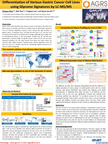
The different cell lines were obviously distinguished through the difference of
relative abundance of various glycan. In overall gastric cancer cell lines, mostly
mannose type glycans dominated and complex type N-glycans were highly
Materials & Method heterogeneous. In distinction from other cell lines, in the KATO-Ⅲ cell line,
Sample Information & Instruments mannose close to the core type significantly increased and external group
containing LacdiNAc was observed. The correlation among GC cell lines was
Established
Name Pathology Lauren type Gender Age Geolocation
year investigated through cluster analysis and the results show a strong relationship
AGS Primary Intestinal F 54 USA 1979 between the N-glycan structure and gastric cancer cell lines. These results
NCI-N87 Metastasis Intestinal M - USA 1976
KATO Ⅲ Primary Diffuse M 15 Japan 1978 suggested that the glycan signatures of each cell line can differentiate GC cell lines.
SNU-1 Primary Diffuse M 44 Korea 1984
Agilent 6530 Summary
SNU-16 Metastasis Diffuse F 33 Korea 1987
Accurate Mass Nano LC-chip Q-
SNU-5 Metastasis Diffuse F 33 Korea 1987 We have worked on cell surface glycan profiling in representative gastric cancer
MKN-45 Primary Diffuse F 62 Japan 1989 TOF MS system
cell lines. Gastric cancer cell lines were successfully profiled using nano LC/MS/MS
MKN-74 Metastasis Intestinal M 37 Japan 1986 : Qualitative profiling
combined with membrane extraction technique. The heterogeneity of each cell
Workflow line was distinguished by moietise of glycans such as fucosylation, sialylation,
LacdiNAc and bisecting structures. These results suggested that the glycan
PNGase F
signatures of each cell line can differentiate GC cell lines. Our findings on GC cell
line-specific glycans may pave the way for accurate diagnosis of heterogeneous
Cells gastric cancer and the potential for molecular target recognition for gastric cancer
Glycoprotein
Cell homogenization Membrane Extraction N-glylcan release LC-MS & LC-MS/MS
with lysis buffer analysis treatment.
We characterized the glycans obtained from representative GC cell lines including Acknowledgement
SNU-1, SNU-16, SNU-5, NCI-N87, AGS, KATO-Ⅲ, MNK-45 and MKN-74. In order to ▪ This work was supported by the Commercialization Promotion Agency for R&D
selectively separate the glycans located on the cell surface, the cell membrane Outcomes of Korea Grant funded by the Korean Government(MSIP) (2022, R&D
extraction was first performed. Subsequently, glycans were enzymatically Equipment Engineer Education Program, 2014R1A6A9064166) & Ministry of
released, enriched and profiled via PGC nanoLC/MS and LC/MS/MS. Using Food and Drug Safety (21172MFDS192-1)
normalized glycan intensity, a correlation among GC cell lines was examined ▪ We thank LABMS members (Laboratory for Advanced Bio-analytical Mass
through hierarchical analysis. Spectrometry) for their tremendous help.
Homepage : www.agrs.kr / E-mail: sugar@cnu.ac.kr

