Page 109 - ebook
P. 109

Blue mussel proteins rescue polystyrene nanoparticles-induced


                                                                                                                                           cellular toxicity in human dermal fibroblasts


                                                                                                    through upregulation of the Nrf2-cytoprotective pathway


                                                                                                                          Choi, Yun-Sik , Goh Eun Hye , and Oh, Yunok *
                                                                                                                                                                                                                                                                                                                                                                  3
                                                                                                                                                                                                                                                                             2
                                                                                                                                                                                               1


                                                      Convergence Research Center for Smart Healthcare, Department of Pharmacology, Kyungsung University, Busan, Korea
                                                   1


                                                      Department of Pharmaceutical Science and Technology, Kyungsung University, Busan, Korea
                                                   2


                                                      Convergence Research Center for Smart Healthcare, Kyungsung University, Busan, Korea
                                                   3


                                                                                                                                                                                                                       Abstract


               With the increased use of plastics, the potential toxicological effects of micro- and nanoplastics on human health have become a global concern. Health and beauty products


               containing microbeads for dermal applications could lead to dermal exposure. But, there is little research that has specifically looked at the toxicity of nanosplastics (NPs) to


               skin penetration. In this study, we investigated the cellular toxicity and skin penetration of polystyrene-amine nanoplastics using Human Dermal Fibroblasts (HDFs). Next, we


               evaluated cytoprotective effects against NPs toxicity in HDFs using blue mussel proteins-derived antioxidant (BMP). BMP showed excellent antioxidant activity in a dose-


               dependent manner, assessed by DPPH assay. Immunostaining and western blot analysis revealed that BMP pre-treatment significantly prevented NPs cellular toxicity by


               inhibiting of mitochondrial ROS generation and caspase-3 activation, and up-regulation of Nrf-2 expression. These results suggest that BMP treatment attenuated cytotoxicity of


               NPs through Nrf2-mediated antioxidant defense mechanism. Therefore, BMP could be considered for use as an antioxidant functional health food against NPs-induced toxicity.


                                                                                                 Introduction                                                                                                                                                                                                                                      Results


                                                                                                                                                                   • Membrane damage

                                                                                                                                                                   • Oxidative stress                                                                                                                                                        A                                                                                                                         B


                                                                                                                                                                   • Immune response


                      Skin contact


                of plastic particles                                                                              Skin penetration
                                                                                                                                                                                                BMP (< 1 kDa)


                        Cytoprotective effects of BMP on PS-NPs-induced cellular toxicity in human dermal fibroblasts


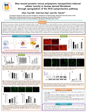
                 Fig. 1. Schematic illustration showing the pathway of human skin exposure to nanoplastics.                                                                                                                                       Fig. 5. The effects of BMP on mitochondrial membrane potential (MMP) loss. (A) MMP disruption


                                                                                                                                                                                                                                                  identified by Mito Tracker                                       TM       Red kit under a fluorescence microscope, and (B) graphical


                                                                                                         Results                                                                                                                                  quantification showing decrease of MMP level on HDF after 24 of treatment with NPs and BMP at


                      A                                                                   B                                                                     C                                                                                 different concentrations. * p < 0.05 vs. control, # p < 0.05 vs NPs only.


                                                                                                                                                                               BMP concentration (g/mL)


               Fig. 2. Antioxidant abilities of blue mussel hydrolysates with various proteolytic enzymes


               determined by (A) DPPH scavenging assay and (B) ORAC assay. (C) DPPH scavenging


               assay of three concentrations of blue mussel hydrolysate by pepsin (BMP). a–d Different                                                                                                                                            Fig. 6. Immunofluorescence staining of cleaved caspase-3 in HDF cells. BMP was treated with


               letters on the bar charts indicated significant differences (p < 0.05). * p < 0.05 vs. control.                                                                                                                                    various concentration (10, 25, and 50 g/mL) with 0.01% NPs in HDF cells.


                                              A                                                                          B

                                                                                                                                                                                                                                                    A                                                                                                                                                           B


               Fig. 3. Cytoprotective effects on human dermal fibroblasts (HDF) after of treatment with (A)


               NPs only and (B) NPs with BMP at different concentrations. The cells were pre-treated with


               various concentration of BMP for 1 h followed by 0.0.1% NPs exposure and 24 h incubation.                                                                                                                                          Fig. 7. Representative western blotting of Nrf-2, and (B) the relative expression level of cleaved


               * p < 0.05 vs. control, # p < 0.05 vs NPs only. BMP was pre-treated before 1 h followed                                                                                                                                            Nrf-2 protein level in NPs-induced cellular damage. Cells were pretreated with BMP for 1 h


              A                                                                                                                                                                                                                                   followed by 0.0.1% NPs exposure and 24 h incubation.


                                                                                                                                                                                                                                                                                                                                                Summary


                                                                                                                                                                                                                                                 • In this study, we demonstrated the anti-atherosclerotic effect of blue mussel hydrolysates by


                                                                                                                                                                                                                                                       pepsin (BMP).

                   B                                                                                                   C                                                                                                                         • The antioxidant effect by BMP was attributed to the promotion of intracellular antioxidant


                                                                                                                                                                                                                                                       defense capacity, such as the inhibition of ROS generation, MMP disruption, caspase-3 activation,


                                                                                                                                                                                                                                                       and upregulation of Nrf-2 expression in NPs-induced HDF damage.


                                                                                                                                                                                                                                                 • Our findings provide new insight of potential use of marine proteins and BMP might considered


                                                                                                                                                                                                                                                       natural antioxidant ingredients for antioxidant functional health food against NPs-induced cellular


                                                                                                                                                                                                                                                       toxicity.


                                                                                                                                                                                                                                                                                                                                              Reference


               Fig. 4. (A) Determination of intracellular ROS production by DCFDH-DA staining under a                                                                                                                                           • Zulueta, A.; Esteve, M.J.; Frígola, A. ORAC and TEAC assays comparison to measure the antioxidant capacity of


               fluorescence microscope and quantitative intracellular DCF fluorescence intensity after 24 of                                                                                                                                          food products. Food Chem. 2009, 114, 310–316.


               treatment with (B) NPs only and (C) NPs with BMP in HDF cells. * p < 0.05 vs. control, # p                                                                                                                                       • Choi, Y.-J.; Kang, J.-S.; Park, J.H.Y.; Lee, Y.-J.; Choi, J.-S.; Kang, Y.-H. Polyphenolic flavonoids differ in their


               < 0.05 vs NPs only                                                                                                                                                                                                                     antiapoptotic efficacy in hydrogen peroxide–treated human vascular endothelial cells. J. Nutr. 2003, 133, 985–991.
   104   105   106   107   108   109   110   111   112   113   114