
Page 43 - I. Chemical biology and drug discovery
P. 43
Berbamine inhibits BDNF-induced angiogenesis of HUVECs in vitro and in vivo
Yu Jin Kim, Jang Mi Han and Hye Jin Jung*
Department of Pharmaceutical Engineering & Biotechnology, Sun Moon University, 70, Sunmoon-ro 221, Tangieong-myeon, Asan-si Chungnam 31460, Korea
ABSTRACT
Brain-derived neurotrophic factor (BDNF), a neurotrophin, plays a critical role in neural development through the activation of its specific receptor, tropomyosin-related
kinase B (TrkB). Recent reports have revealed that BDNF can promote angiogenesis and thus contributes to growth and metastasis of numerous tumor types. Although
berbamine, a natural compound from the plant Berberis amurensis, is known to exert an anticancer activity by targeting Ca /calmodulin-dependent protein kinase II
2+
(CaMKII), its antiangiogenic activity in endothelial cells has been not identified. We here investigated the effect of berbamine on angiogenesis stimulated by BDNF. As a
result, berbamine effectively suppressed the BDNF-induced angiogenic phenotypes such as proliferation, invasion, tube formation, and adhesion of human umbilical
vein endothelial cells (HUVECs) without exhibiting cytotoxicity. In addition, it potently inhibited the neovascularization of chorioallantoic membrane of growing chick
embryo in the presence of BDNF. The antiangiogenic effect of berbamine was also associated with the downregulation of ROS generation increased by BDNF. The
molecular mechanism study to identify the role of BDNF/TrKB/CaMKII axis in the antiangiogenic effect of berbamine is currently underway.
BACKGROUND & AIM METHODS
Angiogenesis is an essential process in tumor growth and metastasis, and controlling it is a promising Cell viability assay
strategy that can prevent cancer progression. Cell proliferation assay
Brain-derived neurotrophic factor (BDNF) is one of the neurotrophic factors, and recent studies have
shown that it contributes to tumor growth and metastasis by promoting angiogenesis. Chemoinvasion assay
Berbamine, a natural compound from plant Berberis amurensis, is known to exert an anticancer effect Capillary tube formation assay
by targeting Ca2+/calmodulin-dependent protein kinase II (CaMKII), but its antiangiogenic activity has Adhesion assay
not been fully identified.
This study evaluates the inhibitory effect of berbamine on the BDNF-induced angiogenesis of human H2DCFDA staining assay
umbilical vein endothelial cells (HUVECs). Chorioallantoic membrane (CAM) assay
RESULTS
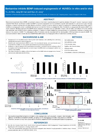
A B C
Chemical structure of Berbamine
BDNF (50ng/ml) - + + + BDNF (50ng/ml) - + + +
Berbamine (μM) - - 10 20 Berbamine (μM) - - 10 20
Berbamine significantly inhibited the BDNF-induced proliferation of HUVECs without exhibiting cytotoxicity.
D E
BDNF (50ng/ml) - + + +
Berbamine (μM) - - 10 20
Berbamine dose-dependently decreased the BDNF-stimulated ROS
production in HUVECs.
F
BDNF (50ng/ml) - + + +
Berbamine (μM) - - 10 20 BDNF (4μg) - + +
Berbamine (10μg) - - +
Berbamine inhibited the BDNF-induced invasion, tube formation, and adhesion Berbamine significantly suppressed the BDNF-induced neovascularization
of HUVECs in a dose-dependent manner. of CAM in growing chick embryos.
CONCLUSION Contact
• Our results showed that berbamine inhibited in vitro angiogenesis, such as growth, invasion, tube formation, and information
adhesion of HUVECs stimulated by BDNF, as well as in vivo angiogenesis of CAM in the presence of BDNF.
• Therefore, berbamine may have a potential to effectively suppress BDNF-mediated pathological angiogenesis.
Yu Jin KIM
REFERENCES Department of
Pharmaceutical Engineering
1. Han JM, et al. Hovenia Dulcis Thunb. And Its Active Compound Ampelopsin Inhibit Angiogenesis Through Suppression of VEGFR2 & Biotechnology, Sun Moon
University
Signaling and HIF-1α Expression. Oncology Reports. 2017.
2. T. Usui, et al. Brain-derived neurotrophic factor promotes angiogenic tube formation through generation of oxidative stress in human petaldew17@naver.com
vascular endothelial cells. ACTA PHYSIOLOGICA. 2014.