


Page 99 - ebook
P. 99
Inhibition of histone deacetylase 1/2 enhanced NK cell mediated

                   anticancer immunity through induction of NKG2D ligands


     Haeryung  Cho 1 , 2  , Eun Jung  Yun 1 , 2 , Jaeho Bae 1 , 2

     Department  of  Biochemistry,  PNU  GRAND  Convergence  Medical  Science  Education  Research  Center,  Pusan  National
     University School of  Medicine,  Yangsan  50612, Korea

     Abstract                          Introduction
     Histone  acetylation  is  an  epigenetic  mechanism  that   Lung cancer is one of the most common cancers and
     regulates the expression of various genes such as the   due to a high relapse rate and distant metastasis has
     natural killer group 2, member D (NKG2D) ligand, and   the highest mortality cancer (1). The important factors
     reduced  by  overpressure  in  cancer  cells.  NKG2D   for  survival  of  lung  cancer  patients  is  the  host's
     ligand  is  a  molecule  that  activates  NKG2D  receptor   immunity  against  cancer  and  the  prognosis  of  lung
     expression in immune cells and is overexpressed by   cancer  patients  can  be  improved  by  enhancing  the
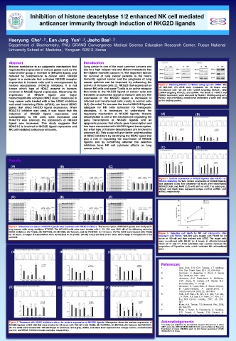
     histone  deacetylases  (HDACs)  inhibitors.  It  is  not   activity of immune cells (2). NKG2D that expressed on   Figure 3. Silencing HDAC1 or HDAC2 using specific siRNA. The
     known  which  type  of  HDAC  enzyme  in  humans   human NK cells and some T cells is an active receptor   (A)  NCI-H23,  (B)  A549  were  incubated  for  48  hours  after
                                                                          transfection  with  150  nM  siH1  (siRNA  targeting  HDAC1),  siH2
     involved in NKG2D ligand expression. Observing  the   that  binds  to  the  NKG2D  ligand  of  cancer  cells  and   (siRNA targeting HDAC2), and scRNA. The levels of HDAC1 and
     expression   of   NKG2D   ligand   and   major   transmits an activation signal to immune cells (3). The   HDAC2 expression were analyzed by Western blotting using anti-
     histocompatibility complex (MHC) class I molecules in   expression  of  the  NKG2D  ligand  is  increased  in   HDAC1 and anti-HDAC2 monoclonal antibodies. β-actin was used
     lung cancer cells treated with a few HDAC inhibitors   infected and transformed cells, rarely in normal cells   as the loading control.
     and small interfering RNAs (siRNA), we found HDAC   (4,5). (In order) To increase the level of NKG2D ligands
     genes  that  affect  NKG2D  ligand  expression.  When   adequate  for  NK  cells  activation  for  therapeutic
     HDAC1/2  inhibitor  was  used,  it  was  found  that  the   strategies,  it  is  necessary  to  understand  the
     induction   of   NKG2D   ligand   expression   and   regulatory  mechanism  of  NKG2D  ligands.  Histone   (A)   (B)
     susceptibility  to  NK  cells  were  increased  and   deacetylation is one of the mechanisms regulating the
     HDAC1/2  was  silenced,  the  expression  of  NKG2D   gene  transcription  of  NKG2D  ligands  and  an
     ligand  was  increased.  This  study  suggests  that   epigenetic process that affects gene transcription and
     HDAC1/2 is involved in NKG2D ligand expression and   has been associated with NKG2D ligand transcription,
     NK cell-mediated anticancer immunity.   but what type of histone deacetylases are involved is
                                       unknown (6). This study will give better understanding
                                       of HDAC inhibitors by identifying the HDAC types that
                                       play  a  role  in  regulating  the  expression  of  NKG2D
                                       ligands  and  by  confirming  whether  the  selective
                                       inhibitors  have  NK  cell  cytotoxic  effects  on  lung
                                       cancer cells (7).                   (C)             (D)
     Results
      (A)                   (B)                    (C)

                                                                          Figure 4. Surface expression of NKG2D ligands after HDAC1 or
                                                                          HDAC2 silencing. Surface protein expression of NKG2D ligands
                                                                          was  analyzed  using  flow  cytometry  48  hours  after  transfecting
                                                                          NCI-H23 (A,B) and A549 (C,D) with siH1 or siH2. The solid gray,
                                                                          dotted,  and  black  lines  represent  isotype  control,  scRNA,  and
                                                                          siRNA, respectively.
      (D)                   (E)                    (F)


     Figure 1. Altered transcription of NKG2D ligands after treatment with HDAC inhibitors. The transcription of NKG2D ligands was analyzed in
     lung cancer cells using multiplex RT-PCR. The NCI-H23 cells were each treated with 1, 10, 100, and 1000 nM of the following selective
     HDAC inhibitors: (A) FK228, (B) RGFP966, (C) MC1568, (D) Tubacin, and (E) PCI34051 for 18 hours. (F) The A549 were treated with FK228   Figure  5.  Selective  cell  death  by  NK  cell  cytotoxicity  after
     for 18 hours. Changes in transcription were normalized to the β-actin (ACTB) and presented as the mean fold change in comparison to the   treatment  with  FK228.  NCI-H23  were  treated  with  FK228  for  24
     controls.
                                                                          hours  at  100  nM  and  then  stained  with  CFSE.  The  target  cells
                                                                          were  co-cultured  with  NK-92  for  4  hours  at  effector-to-target
                                                                          ratios  of  1:1  and  5:1.  Flow  cytometry  was  used  to  measure  the
                                                                          proportion of PI-positive cells, which indicated NK cell-mediated
                                                                          lysis.
      (A)                   (B)                   (C)
                                                                           References
                                                                           1.      Dela  Cruz,  C.S.  Clin.  Tanoue,  L.T.  Matthay,
                                                                                   R.A. Clin. Chest.  Med. 2011, 32, 605–644.
                                                                           2.      Gonzalez,  H.  Hagerling,  C.  Werb,  Z.  Genes
                                                                                   Dev. 2018, 32, 1267–1284.
                                                                           3.      Jamieson,  A.M.  Diefenbach,  A.  McMahon,
                                                                                   C.W.  Xiong,  N.  Carlyle,  J.R.  Raulet,  D.H.
                                                                                   Immunity 2002, 17, 19–29.
     (D)                   (E)                   (F)                       4.      Gonzalez,  S.  Lopez-Soto,  A.  Suarez-Alvarez,
                                                                                   B.  Lopez-Vazquez,  A.  Lopez-Larrea,  C.
                                                                                   Trends Immunol. 2008, 29,  397–403.
                                                                           5.      Park, S.W. Bae, J.H. Kim, S.D. Son, Y.O. Kim,
                                                                                   J.Y. Park, H.J. Lee, C.H. Park, D.Y. Kim, J.Y.
                                                                                   Lee, M.K. Cancer  Investig.  2007,  25,  299–
                                                                                   307.
                                                                           6.      Khan, A.N. Tomasi, T.B. Immunol. Res. 2008,
                                                                                   40,164–178.
                                                                           7.      Bhat,  J.  Dubin,  S.  Dananberg,  A.  Quabius,
                                                                                   E.S.  Fritsch,  J.  Dowds,  C.M.  Saxena,  A.
                                                                                   Chitadze, G. Lettau, M. Kabelitz, D. Immunol.
                                                                                   2019, 10, 569.
     Figure 2. Treatment with HDAC inhibitors alters the surface expression of NKG2D ligands. Histograms show the surface expression of   Acknowledgements
     NKG2D ligands in NCI-H23 that were treated for 24 hours with 100 nM of (A) FK228, (B) RGFP966, (C) MC1568, (D) Tubacin, (E) PCI34051.   This study was supported by the National Research Foundation of Korea
     (F) The A549 were treated with 100 nM FK228 for 24 hours. Solid gray, dotted, and black lines represent the isotype control, media-treated   (NRF) grant (No. NRF-2018R1D1A1B07047437) and the National Research
     control, and HDAC inhibitor-treated samples, respectively.            Foundation  of  Korea  (DIRAMS)  grant  by  the  Korean  government  (MSIP)
                                                                           (50593-2021) of Korea.

