
Page 59 - F. Cell biology
P. 59
Colocalization with MMP-7 in the distal colon is crucial for syndecan-2 shedding during
chronic inflammation
Heejeong Hong¹, Hyun-Kuk Song¹, Bohee Jang¹, Eun-Hye Park¹, Dong-Soo Han², Seong-Eun Kim³, Eok-Soo Oh¹,*
¹Department of Life Sciences and the Research Center for Cellular Homeostasis , Ewha Womans University, Seoul 03760, Republic of Korea, ²Department of Internal Medicine, Hanyang University
College of Medicine, Guri 1923, Republic of Korea, ³Department of Internal Medicine, Ewha Womans University School of Medicine, Seoul 07985, Republic of Korea
ABSTRACT
Previous studies showed that the expression of the cell surface proteoglycan, syndecan-2, was elevated during both chronic inflammation and cancer development, and extracellular domain release of syndecan-2 was observed
in cancer patients. Here, we investigated whether inflammation triggers syndecan-2 shedding. In C57BL/6 mice with DSS-induced colitis, syndecan-2 shedding began to increase after week 12 of chronic inflammation. The
elevation of the shed syndecan-2 level correlated with the increased expression of syndecan-2 and MMP-7 in distal colon tissues, and colocalization of the two molecules in this colon region was a critical factor in stimulating
syndecan-2 shedding. The serum levels of IL-1, IL-6 and IL-17A were increased at week 15 of inflammation. Among them, only IL-6 was altered in trans-distal colon tissues: its mRNA expression level gradually increased from
week 9 to week 15. IL-6 directly induced syndecan-2, MMP-7 expression and syndecan-2 shedding in ex vivo cultured distal colon tissues and adenoma cell lines from the distal colon. Interestingly, hypoxic conditions increased
syndecan-2 expression but attenuated syndecan-2 shedding by suppressing MMP-7 expression. Finally, we observed that the frequency of syndecan-2 and MMP-7 colocalization was increased in the AOM-DSS-induced mouse
model of adenoma and in tissues of (left-sided) colon cancer patients, and that this colocalization was paralleled by increased serum levels of shed syndecan-2. Together, these data demonstrate that IL-6 produced during
chronic inflammation induces syndecan-2 shedding in the distal colon by regulating MMP-7 expression.
INTRODUCTION
The inflammatory response is a defense mechanism that evolved to protect higher organisms from infection and injury (1, 2). Notably, chronic inflammation has long been linked to cancer: If inflammation is chronically
unregulated, there is a high possibility of tumorigenesis and/or tumor progression. About 20% of cancers begin with chronic inflammation, which also promotes tumorigenesis by producing numerous cytokines and chemokines.
Moreover, since cancers induced by chronic inflammation are usually genetically stable, they typically exhibit a high level of cancer drug resistance (3). One mechanism used by the immune system to regulate inflammation is the
shedding (via cleavage of the extracellular domain) of a modulating protein. Since leukocyte recruitment is a key step in basic inflammatory responses, it is critically important to precisely regulate the shedding of adhesion
molecules, such as L-selectin, ICAM-1 and VCAM-1, which contribute to leukocyte recruitment. The extracellular domain of desmoglein-2 is cleaved by the pro-inflammatory cytokines, IL-1β and TNF-α, in the mucosal barrier,
and this shedding may help rebuild the colonic structure after inflammation (4). TNF-α is a well-known pro-inflammatory cytokine that is initially produced as a type II transmembrane protein; when an inflammatory response is
triggered, its ectodomain is cleaved to activate the cytokine. Activated TNF-α has been shown to initiate the TNF-α -dependent cytokine cascade to induce severe skin inflammation (5). Moreover, the shedding of IL-6 receptor α
and HB-EGF was found to occur during lung infection; this boosts the inflammatory response and the ectodomain cleavage of CD44, leading to the recruitment of leukocytes during eosinophilic pneumonia (6, 7). Therefore, the
existing evidence indicates that shedding is a general biological mechanism for the regulation of various inflammation-associated diseases, including cancer.
During cancer progression, the shedding of cadherins is a very common event. The cleavage of P-cadherin is related to breast cancer (8, 9): Soluble P-cadherin (sP-cadherin) was highly detected in nipple aspirate fluids at a
level that depended on the cancerous stage (9), and elevation of sP-cadherin was described as enhancing the migration and invasion of breast cancer (8, 9). Meanwhile, the type III TGF-β receptor (Tβ RIII) was found to exert
different functions before and after its shedding: Tβ RIII itself affects cancer cells in a cancer type-specific manner, whereas soluble Tβ RIII (sTβ RIII) inhibits cellular invasiveness in various types of cancer, such as pancreatic,
non-small cell lung and breast cancer (10–12).
Syndecan is a transmembrane-bounded heparan sulfate proteoglycan that functions as a cell surface receptor to mediate and regulate both inflammation and cancer. Since heparan sulfate binds to various cell surface and matrix
molecules, including cytokines and chemokines, syndecan is able to modulate inflammatory cell maturation, activation and other related functions (13). Interestingly, the extracellular domain shedding of syndecans is also
common during inflammation and cancer. Specially, Syndecan-2 is not expressed in the normal epithelium, but rather exhibits upregulation during chronic inflammation and cancer progression (14, 15). Studies have shown that:
chronic inflammation can induce the expression of colonic syndecan-2 (14); induced syndecan-2 expression in inflammatory macrophages can regulate fibroblast growth factor activity (16); and syndecan-2 expressed on
activated primary human CD4+ lymphocytes can regulate T cell activation (17). These findings suggest that syndecan-2 may also regulate inflammatory responses. Our group further showed that IL-1α induces syndecan-2
shedding in colorectal cancer cell lines (18), and that shed syndecan-2 stimulates colorectal cancer activities (16). However, while the level of shed syndecan-2 is known to be elevated in colorectal cancer, no previous study has
examined shed syndecan-2 in the context of the inflammatory response. Here, we used mouse models of chronic colitis to examine the mechanism responsible for syndecan-2 shedding during inflammation.
RESULTS
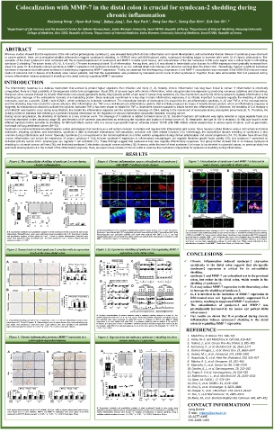
Figure 1. The extracellular shedding of syndecan-2 occurs during Figure 4. Chronic inflammation causes colocalization of syndecan-2 Figure 7. Colocalization of syndecan-2 and MMP-7 is detected in
chronic inflammation. with MMP-7 at distal colonic tissues. tumor tissues, especially in distal colon.
Prox Trans Dist A C57BL/6 IL-17A K/O B RCC LCRC
A Time (d) B Time (d) D Week 9 12 15 9 12 15 9 12 15 Non-tumor Tumor Non-tumor Tumor Non-tumor Tumor Non-tumor Tumor
0 4 10 0 5 10 15 20 25 30 Wks 9 12 15 9 12 15 SDC2 SDC2
DSS H O DSS H O DSS H O DSS H O - SDC2
2
2
2
2
DSS
+ MMP7 MMP7
Vh DSS
Vh DSS
Relative serum SDC2 Relative serum SDC2 a-SDC2 Ponceau.S MMP7 DAPI DAPI
*
Vh DSS
4 7 10 10 20 30 Relative serum SDC2
Time (d) Time (d) DAPI Merge Merge
C Wks 9 12 15
Wks 13 14 15 13 14 15 a b c d
21 Wks 9 12 15 9 12 15 Merge
- -
H 2 O
DSS
1 cycle (6 repeats) 7 + a-SDC2 Ponceau.S DSS + A B C
DSS Vh DSS * a-SDC1 Ponceau.S C Non-tumor Tumor Non-tumor Tumor
LCRC
RCC
Vh DSS
0 Relative serum SDC2 Relative serum SDC1
Time (d) SDC2
Wks 13 14 15
Wks 9 12 15
Double-immunofluorescence staining of inflammation induced colonic tissues at 9, 12 and 15th weeks. A. Double-immunofluorescence staining of SDC-2 and MMP-7 in azoxymethane (AOM)+DSS mouse
A-B. Experiment schedule and quantitative analysis of shed syndecan-2 levels in acute (A) and chronic Syndecan-2 was stained with FITC-conjugated goat anti-mouse antibody, MMP-7 was stained with Texas cancer animal model (Colitis-associated cancer animal model). B. Double-immunofluorescence staining
inflammation 30days(B) mice serum were performed using box-whisker plot. C-D. Experiment schedule Red-conjugated goat anti-rabbit antibody and nuclei were stained with DAPI. A, B, C are high of SDC-2 and MMP-7 in human cancer patient tissue. Syndecan-2 was visualized with FITC-conjugated
and quantitative analysis of shed syndecan-2 and -1 levels in chronic inflammation (15weeks) mice magnification pictures of each white box. White arrows was indicated the colocalization of syndean-2 with goat anti-mouse antibody, MMP-7 was visualized with Texas Red-conjugated goat anti-rabbit antibody
serum were performed using box-whisker plot. The shed syndecan-2 and -1 level in vehicle mice was MMP-7. Scale bars: 50μm, A-C: Scale bars: 100μm. and nuclei were visualized with DAPI. C. SDC-2 immunofluorescences activity difference in non-tumor
used as a control. *p < 0.05
and tumor. a-d are high magnification pictures of each white box. White arrows indicated by the
colocalization of syndean-2 with MMP-7. Scale bars: 50μm, a-d: Scale bars: 100μm.
Figure 2. Serum levels of shed syndecan-2 correlate with its expression Figure 5. IL-6 promotes shedding of syndecan-2 by regulating MMP-7
levels at the trans-distal colon. expression at the distal colon. CONCLUSIONS
Wks Prox Trans Dist
A Vehicle 9 12 15 A 1000 IL-1a Vehicle 8 DSS C 4 * 4 * * Chronic inflammation induced syndecan-2 expression
3
3
IL-1
Prox Trans Dist 500 6 ** Relative Expression SDC2 2 2 * specifically at the distal colon suggests that site-specific
1
1
0
4
0
3
3
6
2
Relative Expression 0 2 * * ** * ** Wks * 12 15 20 9 12 15 Relative shed DSS -IL-1 - +IL-1 + DSS -IL-6 - +IL-6 + + * shedding.
0 MMP7
SDC2 SDC2 4 *** ** * * ** * ** Concentration (pg/ml) 200 IL-6 *** 40 IL-17A *** IL-1 - + - * + IL-6 - + - * + syndecan-2 expression is critical for its extracellular
2
2
**
0
0
1
1
0
2
*
100
-
-
+
0
0
syndecan-2 and MMP-7 are colocalized not in the proximal
SCD2
SDC1
9
1
SDC1 DSS - + - + - + B 4 Prox Trans Dist Tissue Prox Trans Dist Prox Trans Dist Prox Trans Dist Prox Trans Dist colon, but rather in the distal colon, which results in the
0
Wks 9 12 15 IL-1a 3 ** D DSS - + - Distal + shedding of syndecan-2.
2
Proximal
1
4
B Vehicle 9 Wks 15 Relative Expression IL-1 0 ** * * * SDC2 - HCT116 IL-6 IL-17A - SNU1235 IL-17A - DLD1 IL-6 IL-17A - SW480 IL-6 IL-17A IL-6 may induce MMP-7 expression in the descending colon
3
to increase the shedding of syndecan-2.
IL-1a
IL-1a
IL-1a
IL-1a
IL-1
IL-1
IL-1
IL-1
IL-6
2
12
1
0
MMP7
Proximal SDC2 100 Proximal Trans-distal IL-6 4 *** * * ** ** Relative GAPDH * ** * ** 3 ** IL-6 is involved in the induction of MMP-7 expression in
*
3
**
2
3
Expression
DSS-treated mice and hypoxia probably suppressed IL-6
SDC2
2
1
2
80
1
1
0
***
0
0
4
60
secretion, resulting in suppressed MMP-7 expression.
8
*
8
*
IL-17A
MMP-7
*
3
4
4
2
Trans-distal % of expressed area 40 Vehicle 9 12 15 Wks - 9 + - 12 + - 15 + shed SCD2 0 * ** 0 ** The colocalization of syndecan-2 and MMP-7 was
1
0
20
DSS
0
predominantly increased in the mouse and patient distal
a-SDC2
Wks
Ponceau.S colon cancer.
A. Analysis concentrations of various cytokines using a multiplex cytokine analysis in serum. B. The Our results are shown that IL-6 produced during chronic
mRNA expression levels of IL-1α, IL-1β, IL-6, and IL-17A in the proximal, transverse, and distal colon on
A. Immunohistochemistry and qRT-PCR of Syndecan-2 and -1 in chronic inflammation induced mice the indicated days were assessed by qRT-PCR (n=6). C. Analysis of SDC-2 and MMP-7 mRNA inflammation induces syndecan-2 shedding in the distal
colon tissue at 9, 12 and 15 weeks. B. Yellow-colored zone of colonic tissues from each indicated group expression in ex vivo culture and shed SDC-2 in cultured media by with or without IL-1β and IL-6 colon by regulating MMP-7 expression.
and region was syndecan-2 expressed area. Each colored area was calculated and graphically treatment. D. Expression of SDC-2 and MMP-7 mRNA in proximal and distal origin colon cancer cell line
represented compared to vehicle. *p < 0.05, **p < 0.01, ***p < 0.001, Scale bars: 500 μm. by cytokine. shed SDC-2 in cultured media by IL-1α, IL-1β, IL-6, and IL-17A treatment. *p < 0.05, **p
< 0.01, ***p < 0.001
REFERENCE
Figure 3. Chronic inflammation promotes MMP-7 expression in a Figure 6. Hypoxia does not influence syndecan-2 shedding but does 1. Medzhitov, R. Nature 454, 428–435
colon-region-dependent manner. sustain syndecan-2 expression. 2. Kotas, M. E. and Medzhitov, R. Cell 160, 816–827
3. Todoric, J., et al. Cancer Prev Res (Phila). 9, 895–905
4. Kamekura, R., et al. Mol Biol Cell. 26, 3165–3177
A Prox Trans Dist B Prox Trans Dist A Wks Wks B % O 2 - 21 + - 10 + 5. Guinea-Viniegra, J., et al. Genes Dev. 23, 2663–2674
DSS
21 DSS 1 3 9 1 3 9 6. Gomez, M. I., et al. J Immunol. 175, 1930–1936
Vehicle Vehicle H 2 O H 2 O %O 2 21 + - Prox 7. Hayashida, K., et al. Anat Rec (Hoboken). 293, 925–937
1 cycle (3 repeats) 7 10 - + SDC2 Trans 8. Ribeiro, A. S., et al. Oncogene. 29, 392–402
9 9 a-SDC2 Ponceau.S 9. Mannello, F., et al. Cancer Sci. 99, 2160–2169
DSS DSS Dist 10. Gordon, K. J., et al. Carcinogenesis. 29, 252–262
0 21 10 Relative serum SDC2
Wks 12 Wks 12 Time (d) % O 2 10% O 2 - - ++ - - ++ - - ++ Prox 11. Finger, E. C et al. Carcinogenesis. 29, 528–535
DSS - + - + - + - + - + - +
12. Elderbroom, J. L., et al. Mol Biol Cell. 25, 2320–2332
C Wks 1 3 9 MMP-7 Trans 13. Götte, M. FASEB J. 17, 575–591
15 15 900 IL-1α 30 *** 14. Choi, S., et al. FASEB J. 31, 1516–1530
IL-1β
20
600
0
Prox Trans Dist Prox Trans Dist 300 10 Dist 15. Choi, S., et al. Oncotarget. 6, 3874–3886
0
3 MMP-7 ** ** 3 MMP-14 * Concentration (pg/ml) 300 * IL-6 15 ** IL-17A 10 * ** SDC2 6 ** MMP-7 16. Clasper, S., et al. J Biol Chem. 274, 24113–24123
8
*
Relative Expression 2 *** * * Relative Expression 2 * * 100 5 * Relative Expression 6 ** 2 17. Teix, T., et al Mol Immunol. 45, 2905–2919
4
10
200
4
2
1
1
0
0
0
0
DSS
0 0 % O - 21 + - 10 + - 21 + - 10 + DSS - + - + - + - + 18. Kwon, M., et al. Biochem Biophys Res Commun. 446, 487–492
DSS - + - + - + DSS - + - + - + 2 % O 2 21 10 21 10
Wks 9 12 15 Wks 9 12 15
CONTACT INFORMATIOM
A. Experiment schedule and quantitative analysis of shed syndecan-2 levels in mice serum were
A-B. Immunohistochemistry and qRT-PCR of MMP-7(A) and -14(B) in chronic inflammation induced mice performed using box-whisker plot. B. Immunohistochemistry and qRT-PCR of Syndecan-2 and MMP-7 in Jang Bohee
colon tissue at 9, 12 and 15weeks. MMP-7 and MMP-14 expression from each region (n=5) were chronic inflammation induced mice tissues. C. Analysis concentrations of various cytokines using a E mail: bhjang@ewha.ac.kr
analyzed by qRT-PCR (bottom panel). *p < 0.05, **p < 0.01, ***p < 0.001, Scale bars: 50 μm. multiplex cytokine analysis in serum. *p < 0.05, **p < 0.01, ***p < 0.001, Scale bars: 50 μm.
02-3277-4395
010-3268-1450