
Page 67 - F. Cell biology
P. 67
Disrupted-in-schizophrenia 1 enhances the quality of circadian rhythm
by stabilizing BMAL1
Su Been Lee, Jihyun Park, Yongdo Kwak, Young-Un Park, Truong Thi My Nhung, Bo Kyoung Suh, Youngsik Woo, Yeongjun Suh, Eunbyul Cho,
Yubin Won, Tran Diem Nghi, Jinyeong Yoo, Hyeon ah Ji, Sehyung Cho, Sang Ki Park
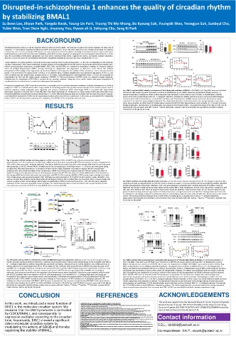
BACKGROUND A GFP-BMAL1 B C D
Flag Flag-DISC1 0.8 0.6 ** MEF(WT) MEF(Disc1 -LI) 2.0 1.5 *
Anticipating the time of day is crucial for organisms that live under recurrent sunlight. The molecular circadian clock system maintains the daily cycle of GFP 95 GFP-BMA L1/tubulin 0.4 BMAL1 72 BMAL1/tubulin 1.0
organisms 1-5. Transcription-translation feedback loop (TTFL) of circadian genes ensures this cycle, which keeps the circadian clock ticking even without Flag 130 0.2 DISC1 95 0.5
external time cues 5. In the mammalian TTFL system, the main component is the heterodimer consisting of Circadian locomotor output cycles kaput tub 55 0.0 tub 55 0.0
Flag-DISC1
(CLOCK) and Brain and muscle Arnt-like protein-1 (BMAL1), which binds to E-box sequences of target genes for transcriptional enhancement 6, 7. In this Flag GFP-BMAL1 WT Disc1-LI
way, the CLOCK/BMAL1 heterodimer upregulates core clock genes, including Period (PERs), Cryptochrome (CRYs), and Nuclear receptor subfamily 1
(Nr1d1) 5. In turn, the action of CLOCK/BMAL1 heterodimer is negatively regulated by PERs and CRYs, thus completing the TTFL 8, 9. E F
MEF(WT) MEF(Disc1 -LI) 1.5 ** *
12 16 20 24 28 32 12 16 20 24 28 32 dexa.(h)
Timed regulation of circadian proteins is essential to the proper working of the circadian timing system. To this end, the degradation of the molecular 1.0
circadian components is vital. Newly synthesized circadian proteins must be degraded at the right time for the accurate pacing of the circadian clock. BMAL1 72 Relative BMA L1/tubulin
Indeed, several circadian components, including BMAL1, PERs, CRYs, and REV-ERBs, are regulated by degradation pathways 10. Glycogen synthase tub 0.5 WT
kinase 3β (GSK3β) phosphorylates BMAL1, thereby enhancing its ubiquitination and degradation 11. Moreover, Casein kinases Iδ/ε (CKIδ/ε) 55 0.0 Disc1-LI
phosphorylate PERs leading to their degradation 10. Chemical inhibition of CKI augments the stability of PERs and induces a more extended circadian 12 16 20 24 28 32 dexa. (h)
period. F-box and leucine-rich repeat protein 3 (FBXL3), an E3 ubiquitin ligase, mediates ubiquitination and subsequent degradation of CRYs 12, and G H I J
FBXL3-mutated mice also exhibit longer circadian period 13. REV-ERBα is phosphorylated by Cyclin-dependent kinase 1 (CDK1) and recognized for 0.0008 0.0003 0.006
ubiquitination by F-box and WD repeat domain containing 7 (FBXW7) 14. The loss-of-function of FBXW7 results in a damped circadian amplitude, Per1 # # Per2 * Cry1 0.003 ** ** Bmal1
indicating that the degradation of REV-ERBα is essential for the circadian amplitude. Altogether, these studies indicate that proper degradation of 0.0006 0.0002 # # 0.004 0.002 ** * *
circadian proteins is a critical factor to maintain circadian period and amplitude. mRNA/GAPDH 0.0004 0.0001 0.002
0.0002
W T WT WT 0.001 W T
Disrupted-in-schizophrenia 1 (DISC1) was first reported as a responsible gene for prevalent psychiatric conditions, including schizophrenia, in a Scottish 0.0000 Disc1 -LI 0.0000 Disc1 -LI 0.000 Disc1 -LI 0.000 Disc1 -LI
pedigree 15. DISC1 is a scaffold protein with a large number of interacting partners that perform various functions in the nervous system, such as 12 16 20 24 28 32 36 40 12 16 20 24 28 32 36 40 12 16 20 24 28 32 36 40 12 16 20 24 28 32 36 40 dexa.(h)
neuronal migration, neurite outgrowth, spine regulation, and synapse maintenance 16-19. Interestingly, DISC1 is associated with sleep-related Fig. 3 DISC1 regulates BMAL1 stability and knockout of Disc1 damps the oscillation of BMAL1. a GFP-BMAL1 and Flag-DISC1 were co-transfected
phenotypes. Expression of human DISC1 in fruit flies alters their sleep homeostasis 20. Furthermore, a mouse model of DISC1 gain-of-function shows into HEK293 cells. Empty Flag vector was used as a control. b Quantification of GFP-BMAL1 levels from (a) relative to tubulin control (n = 5,
increased wakefulness and decreased REM and NREM sleep 21. A close relationship between sleep and circadian clock has been proposed 22; however, biological replicates). c Endogenous BMAL1 protein expression was decreased in Disc1 knockout (Disc1-LI) MEFs. d Quantification of BMAL1
a potential direct involvement of DISC1 in the mammalian circadian system has not been explored yet. In this study, we investigated the relationship protein levels from (c) with tubulin control (n = 5, biological replicates). e Endogenous BMAL1 protein levels were measured in MEFs following
between DISC1 and circadian system and proposed a modulatory role of DISC1 in the mammalian molecular clock. treatment with 1 μM of dexamethasone to synchronize circadian cycle. f Quantification of BMAL1 protein levels from (e) relative to tubulin
control (n = 3 for each time point, biological replicates). The relative band intensities at each time point to the average intensity of entire time
RESULTS points were presented. g-j mRNA levels of circadian genes measured by qRT-PCR after dexamethasone synchronization (n = 3 for each time point,
biological replicates). The expression of circadian genes was reduced in Disc1 knockout MEFs. The panels show levels of Per1 (g), Per2 (h), Cry1 (i),
and Bmal1 (j). Data are means with SEM. *p≤0.05, **p≤0.01, #p≤0.0001, unpaired t-test for (b) and (d); two-way ANOVA and Sidak’s multiple
comparisons for (f), (g), (h), (i) and (j).
A B
2.5 2.0 mDisc1 1.5 A 3500 3000 2500 WT Disc1-LI B 39 38 WT Disc1-LI C 4000 WT Disc1-LI
Relative mRNA/Gapdh 1.5 1.0 Relative mRNA/Gapdh 1.0 0.5 WRA(Turns/1hr) 2000 1500 1000 500 0 0 6 12 18 24 BT( ℃) 37 36 35 34 0 6 12 18 24 HCA(counts/1hr) 3000 2000 1000 0
6
12
****
0.0 0.5 0.0 mDisc1 mPer1 mPer2 mCry1 30000 Zeitgeber Time (hour) 37.0 Zeitgeber Time (hour) 60000 0 Zeitgeber Time (hour) 18 24
****
****
12 16 20 24 28 32 36 40 0 4 8 12 16 20 20000 36.5 40000
After Dexamethasone (h) Circadian Time (h) WRA(Turns/1day) ℃ 1day mean) 36.0 HCA(counts/1day)
C D 10000 BT( 35.5 35.0 20000
1.5 0 WT Disc1-LI 0 WT Disc1-LI 0 WT Disc1-LI
Relative DISC1/tubulin *
CT(h) 0 4 8 12 16 20 KO 1.0 D E
DISC1 95 24.0
tubulin 55 0.5 23.6 23.70
23.70
23.53
0.0 mDISC1 D/D Free Running Period 23.2 23.53
0 4 8 12 16 20
Circadian Time (h)
0
Fig. 1 Expression of DISC1 exhibits oscillating pattern. a mRNA expression of Disc1 in NIH3T3 cells. Cells were treated with 1 μM of WT Disc1-LI
12
dexamethasone for 2-hr to synchronize circadian cycle. mRNA levels of Disc1 were analyzed by qRT-PCR and compared to those of Gapdh (n=5 for CT 0 6 12 WT 18 0 6 12 CT 0 6 Disc1-LI 18 0 6 12
12hr, n=6 for other time points, biological replicates). The relative mRNA levels at each time point to the average mRNA level of entire time points F
were presented. p=0.0126, one-way ANOVA. b mRNA level of Disc1 in hippocampus of wild type mice (n = 3, 3 mice for each time point) during 0.0015 * 0.003 * 0.0020 **
circadian time. Mice were entrained on 12-hr light/12-hr dark cycle for a week; thereafter, mice were in constant dark for a day and were 0.0010 0.002 0.0015
sacrificed under dim red light according to their circadian time. mRNA levels of Disc1 were analyzed using qRT-PCR and compared to those of Per2 level/Gapdh Cry1 level/Gapdh Bmal1 level/Gapdh 0.0010
Gapdh. To compare the expression patterns of circadian genes measured by qRT-PCR each other, the relative mRNA levels at each time point to 0.0005 0.001 0.0005
the average mRNA level of entire time points were presented. p=0.0037 for Disc1, one-way ANOVA. c DISC1 protein levels according to circadian
time in hippocampus were assessed by western blot. Wild type mice were entrained on 12-hr light/12-hr dark cycle for a week; thereafter, mice 0.0000 WT Disc1-LI 0.000 WT Disc1-LI 0.0000 WT Disc1-LI
were on constant dark condition and sacrificed under dim red light according to their circadian time. Disc1 knockout (Disc1-LI mouse) sample was CT16 CT16 CT16
used as a negative control. Arrow indicates DISC1 bands. d Quantification of DISC1 protein level from (c) (n = 4 for each time point, biological Fig. 4 Disc1 knockout mice exhibit altered circadian behaviors. a-c Overall circadian behaviors during L/D cycle (n = 10, biological replicates). Mice
replicates). Protein levels were quantified relative to tubulin. The relative band intensities at each time point to the average intensity of entire were fully entrained to a light/dark (L/D; 12-hr/12-hr) cycle for at least two weeks. For the next 10 days with the same lighting schedule, light/dark
time points were presented. p= 0.0377, one-way ANOVA. Data are means with SEM. profiles were generated as described in Methods. Then, mice were released to a dark/dark (D/D; constant dark) cycle for another 10 days to
determine the internal circadian period. (a) WRA, wheel running activity; (b) BT, body temperature; (c) HCA, home cage activity. Compared to wild
type mice, Disc1 knockout mice (Disc1-LI) showed reduced values during the nighttime. Total measured values per one day were depicted as bar
graphs for wheel running and home cage activities. For body temperature, averaged values were used. d, e Free-running periods were measured
A DISC1 promoter Luc B Distal DISC1 promoter Proximal for wild type (n = 16, biological replicates) and Disc1 knockout (n = 10, biological replicates) mice under the D/D cycle. Disc1 knockout mice showed
Middle
TSS -982 -624 -324 TSS +47 shorter Free-running period than wild type mice. f mRNA levels of circadian genes at circadian time of 16hr (n = 5, biological replicates).
12 Hippocampus was dissected from wild type or Disc1 knockout mice. Data are means with SEM. *p≤0.05, **p≤0.01, ****p≤0.0001, unpaired t-test
**** Distal Luc **** for (a), (b), (c), (e) and (f).
10
Normalized RLA 8 6 4 n.s. Middle Luc * A B input(2.5%) IP: GFP C input(2.5%) IP: myc
HA-ub
HA-ub
CLOCK & BMAL1
0 2 Proximal Luc n.s. None input(2.5%) IP: Flag GFP-BMAL1 + + + + + + + + + + myc-BMAL1 + + + + + + + + + +
shCTL
CLOCK - + + 0 5 10 15 20 Flag-DISC1 + + Flag-DISC1 + + shDISC1 + +
BMAL1 - - + Normalized RLA
GFP-BMAL1 + + + + HA HA
130
Flag 170 170
C None E ChIP primer sets 95 130 130
DISC1 promoter Luc CLOCK & BMAL1 GFP 95 95 95
72
TSS GFP
E-box(-718) E-box(-668) 95
DISC1 promoter Luc **** 10 **** Flag 130 BMAL1 72
TSS **
-718 mutant 8 ****
DISC1 promoter Luc **** D E MEF(WT) MEF(Disc1-LI)
TSS
-668 mutant 6 input(2.5%) IP: GFP 12 16 20 24 28 32 12 16 20 24 28 32 dexa. (h)
DISC1 promoter Luc Precipitated relative to input (normalized) 4 **** **** HA-ub + + + + + + GSK3β p-Y216
TSS GFP-BMAL1 + + + + + + + + 43
-718 -668 mutants myc-GSK3 β S9A + + + + GSK3β 43
0 1 2 3 4 2 Flag-DISC1 + +
Normalized RLA 0 HA DISC1 95
D CLOCK - - + + - - + + 170 tubulin 55
BMAL1 - + - + - + - + 130 F G
None 95
Distal Luc CLOCK & BMAL1 E-box(-718) E-box(-668) 1.5 1.5 2.0
GFP 1.5 *
IP: GFP ab 95 55 1.0 1.0
Distal Luc **** myc 130 Relative p-Y216 /GSK3 Relative DISC1/tubulin Relative p-Y216 /GSK3 1.0
Flag 0.5 0.5
n.s.
-718 -668 mutants GSK3βY216P 0.5 MEF(WT)
DISC1
Normalized RLA 0.0 12 16 20 24 28 32 0.0 0.0 12 16 20 24 28 MEF(Disc1-LI) 32
dexa. (h) dexa. (h)
Fig. 2 Promoter activity of DISC1 is enhanced by CLOCK and BMAL1 through E-box sequences. a DISC1 promoter was cloned into pGL3 vector to Fig. 5 DISC1 inhibits BMAL1 ubiquitination in association with suppression of Y216 phosphorylation of GSK3β. a Co-immunoprecipitation of
monitor promoter activity by luciferase expression. Luciferase assay was performed in HEK293 cells overexpressing CLOCK or together with BMAL1 DISC1 and BMAL1. Flag-DISC1 and GFP-BMAL1 were transfected in HEK293 cells and precipitated with Flag-DISC1. b Ubiquitination assay for
(n = 3, biological replicates). b Luciferase assay with DISC1 promoter sub-regions: distal part (-982 to -624 bp relative to TSS), middle part (-623 to - BMAL1. HA-ubiquitin was transfected in HEK293 cells to assess the ubiquitination of BMAL1. GFP-BMAL1 was transfected with or without Flag-
324 bp relative to TSS), and proximal part (-323 to +47 bp relative to TSS). Luciferase assay was performed in HEK293 cells overexpressing CLOCK DISC1. c Ubiquitination assay of BMAL1 with DISC1 knockdown. HA-ubiquitin was transfected in HEK293 cells to assess the ubiquitination of
and BMAL1 (n = 3, biological replicates). Each group was normalized to the respective control (empty vectors were transfected). c Luciferase assay BMAL1. myc-BMAL1 was transfected with shCTL or shDISC1 for knockdown of DISC1. d Ubiquitination assay of BMAL1 with GSK3β S9A and DISC1.
with E-box-mutated DISC1 promoter. Luciferase assay was performed in HEK293 cells overexpressing CLOCK and BMAL1 (n = 3, biological HA-ubiquitin was transfected in HEK293 cells to assess the ubiquitination of BMAL1. GFP-BMAL1 was transfected with myc-GSK3β CA and Flag-
replicates). Each group was normalized to the respective control (empty vectors were transfected). d Luciferase assay conducted using the distal DISC1 according to each combination of constructs. e Western blot analysis of Y216 phosphorylation of GSK3β in wild type and Disc1 knockout
part of DISC1 promoter with E-box mutations in HEK293 cells overexpressing CLOCK and BMAL1 (n = 3, biological replicates). Each group was (Disc1-LI) MEFs. MEFs were treated with 1 μM of dexamethasone to synchronize the circadian cycle. Samples were collected from same
normalized to the respective control (empty vectors were transfected). e Chromatin immunoprecipitation (ChIP) was performed in HEK293 cells experimental preparation and western blot was conducted in parallel with the same amount of samples. f Quantification of DISC1 and Y216
transfected with CLOCK or BMAL1 or both to assess direct binding on E-box sequences of DISC1 promoter (n = 3, biological replicates). GFP phosphorylation of GSK3β (n = 6, n = 3 for DISC1, biological replicates). Relative intensities of DISC1 and p-Y216 GSK3β relative to tubulin and total
antibody was utilized to pull down GFP-BMAL1. qPCR was used to quantify the E-box sequences precipitated by GFP-BMAL1. Red arrows indicate GSK3β, respectively, were subjected to analyses. The relative band intensities at each time point to the average intensity of entire time points
estimated primer binding sites for qPCR. For CLOCK and BMAL1 transfection, RFP-CLOCK-myc and GFP-BMAL1 constructs were used for (a), (b), (c), were presented. g Quantification of Y216 phosphorylation levels in wild type and Disc1 knockout MEFs (n = 6, biological replicates). The intensity
(d) and (e). RLA represents relative luciferase activity. Data are means with SEM. *p≤0.05, **p≤0.01, ***p≤0.001, ****p≤0.0001, one-way ANOVA, of p-Y216 GSK3β relative to total GSK3β were subjected to analyses. The relative band intensities at each time point to the average intensity of
Tukey’s post-hoc test for (a), (b), (c), (d) and (e). entire time points were presented. Data are means with SEM. *p≤0.05, two-way ANOVA and Sidak’s multiple comparisons for (g).
CONCLUSION REFERENCES ACKNOWLEDGEMENTS
In this work, we introduced a novel function of 1. Young MW, Kay SA. Time zones: a comparative genetics of circadian clocks. Nat Rev Genet 2001; 2(9): 702-715. This work was supported by the Advanced Research Center Program (Organelle
Network Research Center, 2017R1A5A1015366) and the Original Technology
2. Doherty CJ, Kay SA. Circadian control of global gene expression patterns. AnnuRev Genet 2010; 44(1): 419-444.
DISC1 in the molecular circadian system. We 3. Bell-Pedersen D, CassoneVM, Earnest DJ, Golden SS, Hardin PE, Thomas TL et al. Circadian rhythms from multiple oscillators: lessons from diverse organisms. Nat Rev Genet 2005; 6(7): 544-556. Research Program (2017M3C7A1047875, NRF-2020M3E5E2039894) funded by
4. Dunlap JC. Molecular Bases for Circadian Clocks. Cell 1999; 96(2): 271-290.
5. Takahashi JS. Transcriptional architecture of the mammalian circadian clock. Nat Rev Genet 2017; 18(3): 164-179.
revealed that the DISC1 promoter is controlled 6. Gekakis N, Staknis D, Nguyen HB, Davis FC, Wilsbacher LD, King DP et al. Role of the CLOCK protein in the mammalian circadian mechanism. Science 1998; 280(5369): 1564-1569. Korean National Research Foundation (S.K.P).
7. King DP, Zhao Y, Sangoram AM, Wilsbacher LD, Tanaka M, Antoch MP et al. Positional Cloning of the Mouse Circadian Clock Gene. Cell 1997; 89(4): 641-653.
8. KumeK, Zylka MJ, Sriram S, Shearman LP, Weaver DR, Jin X et al. mCRY1 and mCRY2 are essential components of the negative limb of the circadian clock feedback loop. Cell 1999; 98(2): 193-205.
by CLOCK/BMAL1, and consequently its 9. Shearman LP, Sriram S, Weaver DR, Maywood ES, Chaves I, Zheng B et al. Interacting Molecular Loops in the Mammalian Circadian Clock. Science 2000; 288(5468): 1013-1019.
10. Hirano A, Fu YH, Ptacek LJ. The intricate dance of post-translational modifications in the rhythm of life. Nat Struct Mol Biol 2016; 23(12): 1053-1060.
11. Sahar S, Zocchi L, Kinoshita C, Borrelli E, Sassone-Corsi P. Regulation of BMAL1 Protein Stability and Circadian Function by GSK3 beta-Mediated Phosphorylation. Plos One 2010; 5(1).
12. Busino L, Bassermann F, Maiolica A, Lee C, Nolan PM, GodinhoSIH et al. SCF Fbxl3 Controls the Oscillation of the Circadian Clock by Directing the Degradation of Cryptochrome Proteins. Science 2007;
expression oscillates according to the circadian 15. Millar JK, Wilson-Annan JC, Anderson S, Christie S, Taylor MS, Semple CA et al. Disruption of two novel genes by a translocation co-segregating with schizophrenia. Hum Mol Genet 2000; 9(9): 1415-1423. Contact information
316(5826): 900-904.
13. GodinhoSIH, Maywood ES, Shaw L, Tucci V, Barnard AR, Busino L et al. The After-Hours Mutant Reveals a Role for Fbxl3 in Determining Mammalian Circadian Period. Science 2007; 316(5826): 897-900.
14. Zhao X, Hirota T, Han X, Cho H, Chong L-W, Lamia K et al. Circadian Amplitude Regulation via FBXW7-Targeted REV-ERBα Degradation. Cell 2016; 165(7): 1644-1657.
time. Importantly, DISC1 showed a significant 16. Brandon NJ, Sawa A. Linking neurodevelopmental and synaptic theories of mental illness through DISC1. Nature Reviews Neuroscience 2011; 12: 707.
17. Ishizuka K, Paek M, Kamiya A, Sawa A. A review of Disrupted-In-Schizophrenia-1 (DISC1): neurodevelopment, cognition, and mental conditions. Biol Psychiatry 2006; 59(12): 1189-1197.
role in molecular circadian system by 18. Wilkinson B, Evgrafov O, Zheng D, Hartel N, Knowles JA, Graham NA et al. Endogenous Cell Type-Specific DISC1 Interactomes Reveal Protein Networks Associated to Neurodevelopmental Disorders. S.B.L., lsb0202@postech.ac.kr
Biological Psychiatry 2018.
19. Camargo LM, Collura V, Rain JC, Mizuguchi K, Hermjakob H, Kerrien S et al. Disrupted in Schizophrenia 1 Interactome: evidence for the close connectivity of risk genes and a potential synaptic basis for
modulating the activity of GSK3β and thereby schizophrenia. Molecular Psychiatry 2006; 12: 74.
20. Sawamura N, Ando T, Maruyama Y, Fujimuro M, Mochizuki H, Honjo K et al. Nuclear DISC1 regulates CRE-mediated gene transcription and sleep homeostasis in the fruit fly. Molecular psychiatry 2008;
13(12): 1138-1069.
regulating the stability of BMAL1. 21. Jaaro-Peled H, Altimus C, LeGates T, Cash-Padgett T, ZoubovskyS, Hikida T et al. Abnormal wake/sleep pattern in a novel gain-of-function model of DISC1. Neuroscience research 2016; 112: 63-69. Correspondence: S.K.P., skpark@postech.ac.kr
22. Wulff K, Porcheret K, Cussans E, Foster RG. Sleep and circadian rhythm disturbances: multiple genes and multiple phenotypes. Current Opinion in Genetics & Development 2009; 19(3): 237-246.