
Page 3 - E. Cell adhesion and cytoskeletal dynamics
P. 3
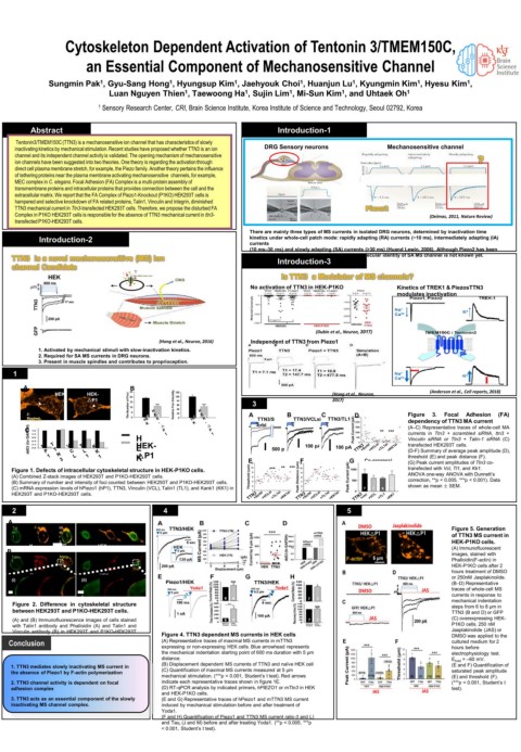
Cytoskeleton Dependent Activation of Tentonin 3/TMEM150C,

                   an Essential Component of Mechanosensitive Channel

           Sungmin Pak , Gyu-Sang Hong , Hyungsup Kim , Jaehyouk Choi , Huanjun Lu , Kyungmin Kim , Hyesu Kim ,
                                                                                        1
                                                               1
                                                                          1
                                                  1
                                    1
                      1
                                                                                                  1
                        Luan Nguyen Thien , Taewoong Ha , Sujin Lim , Mi-Sun Kim , and Uhtaek Oh 1
                                                             1
                                       1
                                                    1
                                                                        1
                     1  Sensory Research Center, CRI, Brain Science Institute, Korea Institute of Science and Technology, Seoul 02792, Korea
       Abstract                                            Introduction-1
    Tentonin3/TMEM150C (TTN3) is a mechanosensitive ion channel that has characteristics of slowly
    inactivating kinetics by mechanical stimulation. Recent studies have proposed whether TTN3 is an ion   DRG Sensory neurons                                   Mechanosensitive channel
    channel and its independent channel activity is validated. The opening mechanism of mechanosensitive
    ion channels have been suggested into two theories. One theory is regarding the activation through   ?
    direct cell plasma membrane stretch, for example, the Piezo family. Another theory pertains the influence
    of tethering proteins near the plasma membrane activating mechanosensitive  channels, for example,
    MEC complex in C. elegans. Focal Adhesion (FA) Complex is a multi-protein assembly of
    transmembrane proteins and intracellular proteins that provides connection between the cell and the
    extracellular matrix. We report that the FA Complex of Piezo1-Knockout (P1KO) HEK293T cells is
    hampered and selective knockdown of FA related proteins, Talin1, Vinculin and Integrin, diminished
    TTN3 mechanical current in Ttn3-transfected HEK293T cells. Therefore, we propose the disturbed FA   Piezo2
    Complex in P1KO HEK293T cells is responsible for the absence of TTN3 mechanical current in ttn3-  (Delmas, 2011, Nature Review)
    transfected P1KO-HEK293T cells.
                                                     There are mainly three types of MS currents in isolated DRG neurons, determined by inactivation time
         Introduction-2                              kinetics under whole-cell patch mode: rapidly adapting (RA) currents (~10 ms), intermediately adapting (IA)
                                                     currents
                                                     (10 ms–30 ms) and slowly adapting (SA) currents (>30 ms) (Huand Lewin, 2006). Although Piezo2 has been
   TTN3  is a novel mechanosensitive (MS) ion        discovered as a RA MS channel (Coste, 2010), molecular identity of SA MS channel is not known yet.
                                                           Introduction-3
   channel Candidate
                                                           Is TTN3  a Modulator of MS channels?
                                                     No activation of TTN3 in HEK-P1KO  Kinetics of TREK1 & PiezosTTN3
                                                                                   modulates inactivation
                                                                  (Dubin et al., Neuron, 2017)
                                  (Hong et al., Neuron, 2016)  Independent of TTN3 from Piezo1
         1. Activated by mechanical stimuli with slow-inactivation kinetics.
         2. Required for SA MS currents in DRG neurons.
         3. Present in muscle spindles and contributes to proprioception.
    1
      A                     B
            HEK    HEK-                                               (Hong et al., Neuron,   (Anderson et al., Cell reports, 2018)
                   △P1                               3                2017)
      Z-                       4  3      4  3        A      B       C      D          Figure  3.  Focal  Adhesion  (FA)
      Stacked                  3  6      3  6         TTN3/S  600  TTN3/VCLsi  600  TTN3/TL1si  dependency of TTN3 MA current
      F-actin
       ddCt (to GAPDH)       H                           500 pA  100 pA  100 pA       Vinculin siRNA or Ttn3 + Talin-1 siRNA (C)
      C                                               600  ms crbl  ms  ms      **  ***  (A–C) Representative traces of whole-cell MA
                                                                                      currents in Ttn3 + scrambled siRNA, ttn3 +
                             HEK-
                                                                                      transfected HEK293T cells.
                             E
                                                                                      (D-F) Summary of average peak amplitude (D),
                             △P1
                             K
                                                                                      (G) Peak current amplitudes of Ttn3 co-
                                                                             on
                                                                                      transfected with Vcl, Tl1, and Kk1.
   Figure 1. Defects of intracellular cytoskeletal structure in HEK-P1KO cells.  E  ***  **  ***  F  G  Co-expressi  threshold (E) and peak distance (F).
   (A) Combined Z-stack images of HEK293T and P1KO-HEK293T cells.                     ANOVA one-way ANOVA with Dunnett’s
   (B) Summary of number and intensity of foci counted between HEK293T and P1KO-HEK293T cells.  correction, **p < 0.005, ***p < 0.001). Data
   (C) mRNA expression levels of hPiezo1 (hP1), TTN3, Vinculin (VCL), Talin1 (TL1), and Kank1 (KK1) in   shown as mean ± SEM.
   HEK293T and P1KO-HEK293T cells.
   2                               4                                     5
   A
   Tal  F-actin  M                                                                             Figure 5. Generation
   in-       e                                                                                 of TTN3 MS current in
   1         r
             g                                                                                 HEK-P1KO cells.
       H     10 e μm  HEK-                                                                     (A) Immunofluorescent
  B    E             △P1                                                                       images, stained with
   Talin-  K Vincu  M Merg
   Talin
        Vinc
   1    lin  e                                                                                 Phalloidin(F-actin) in
        ulin  er                                                                               HEK-P1KO cells after 2
             g                                                                                 hours treatment of DMSO
       H     e        HEK-
   (i)  E         (ii)  △P1                                                                    or 250nM Jasplakinolide.
       K                                                                                       (B–D) Representative
                                                                                               traces of whole-cell MS
                                                                                               currents in response to
   2 μm           2 μm                                                                         mechanical indentation
   Figure 2. Difference in cytoskeletal structure                                              steps from 6 to 8 μm in
   between HEK293T and P1KO-HEK293T cells.                                                     TTN3 (B and D) or GFP
   (A) and (B) Immunofluorescence images of cells stained                                      (C) overexpressing HEK-
   with Talin1 antibody and Phalloidin (A) and Talin1 and                                      P1KO cells. 250 nM
   Vinculin antibody (B) in HEK293T and P1KO-HEK293T  Figure 4. TTN3 dependent MS currents in HEK cells  Jasplakinolide (JAS) or
   cells.                                                                                      DMSO was applied to the
   Conclusion                     (A) Representative traces of maximal MS currents in mTTN3    cultured medium for 2
                                  expressing or non-expressing HEK cells. Blue arrowhead represents   hours before
                                  the mechanical indentation starting point of 600 ms duration with 5 μm   electrophysiology test.
                                  distance.                                                    E hold = –60 mV.
   1. TTN3 mediates slowly inactivating MS current in   (B) Displacement dependent MS currents of TTN3 and naïve HEK cell  (E and F) Quantification of
   the absence of Piezo1 by F-actin polymerization   (C) Quantification of maximal MS currents measured at 5 μm   saturated peak amplitude
                                  mechanical stimulation. (***p < 0.001, Student’s t test). Red arrows   (E) and threshold (F).
   2. TTN3 channel activity is dependent on focal   indicate each representative traces shown in figure 1E.  (***p < 0.001, Student’s t
   adhesion complex               (D) RT-qPCR analysis by indicated primers, hPIEZO1 or mTtn3 in HEK   test).
                                  and HEK-P1KO cells.
   3. TTN3 acts as an essential component of the slowly   (E and G) Representative traces of hPiezo1 and mTTN3 MS current
   inactivating MS channel complex.  induced by mechanical stimulation before and after treatment of
                                  Yoda1.
                                  (F and H) Quantification of Piezo1 and TTN3 MS current ratio (I and L)
                                  and Tau i (J and M) before and after treating Yoda1. (**p < 0.005, ***p
                                  < 0.001, Student’s t test).