
Page 1 - E. Cell adhesion and cytoskeletal dynamics
P. 1
Three Dimensional Monolayer Culture of Epithelial and Endothelial Cells on Poly(vinyl alcohol)
Nanofibrous Membrane Containing Integrin-binding Ligands
1
Min-Ho Choi , Yong-Su Kim , Jung-In Shin , Ji-Hyun Lee , Perry Ayn Mayson A. Maza , Minsook Ryu , Jong-Yong Kwak 1,4,5
1,2
1,2
3
1,2
1,2
1 Department of Pharmacology, School of Medicine, Ajou University Suwon 16499, South Korea, 2 Department of Biomedical Sciences, Graduate school, Ajou University Suwon 16499, South Korea, 3 Department of
Allergy, Ajou University School of Medicine, Suwon 16499, South Korea, 4 3D Immune System Imaging Core Center, Ajou University, Suwon 16499, South Korea Korea, 5 Immune Network Pioneer Researcg Center, Ajou
University, Suwon 16499, South Korea, 6 Nanofaentech Inc. Kimhae 50969, South Korea
BACKGROUND AIM
Adherence of epithelial cells to traditional two-dimensional (2D) culture dish may induce aberrant In this study, we developed a three dimensional (3D) culture system of epithelial and
cell functions, including epithelial-mesenchymal transition. Poly(vinyl alcohol) (PVA) is one of the endothelial cells with PVA nanofibrous membrane which contains integrin-binding
most prevalent and versatile synthetic polymers abundantly used in tissue engineering as peptides to improve cell adhesion, growth, and functions.
biomaterials, but high hydrophilicity of PVA results in poor cell adhesion, leading to aggregation
and spheroid formation of cultured cells on PVA nanofibrous membrane.
METHODS
1. Electrospinning - Electrospun nanofibers were prepared from blends of PVA, integrin-binding peptides, and chemical cross linkers. The electrospun PVA nanofibrous membranes were then
further crosslinked by exposure of the membranes to HCl vapor and dimethylformamide solution.
2. Cell culture – MLE-12 lung epithelial cells, bEND.3 endothelial cells, mouse primary hepatocytes, CT26 colon cancer cells, HepG2 hepatoma cells, and fibroblasts were plated on 2D culture
dish, PVA nanofibrous membrane, and PVA nanofibrous membrane containing peptide of fibronectin and laminin.
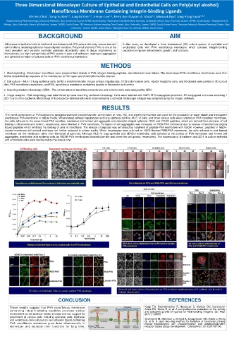
3. Scanning electron microscopy (SEM) - The ultrastructure of nanofibrous membrane and cultured cells were analysed by SEM.
4. Image analysis - Cell morphology was determined by laser scanning confocal microscopy. Cells were labelled with DAPI, FITC-conjugated phalloidin, PE-conjugated anti-zona occluding 1
(ZO-1) and anti-E-cadherin. Morphology of fluorescence-labelled cells were observed using a confocal microscope. Images was analysed using the ImageJ software.
RESULTS
The novel combination of PVA/polyacrylic acid/glutaraldehyde crosslinked with combination of heat, HCl, and dimethylformamide was used for the production of water-stable and transparent
electrospun PVA membrane in culture media. When mouse primary hepatocytes and lung epithelial cell line, MLE-12 cells, and other various cells were cultured on PVA nanofiber membrane,
the cells adhered to the cross-linked PVA nanofiber membrane but formed cell aggregate and discoidal shaped spheroid. RGD and YIGSR peptides, which are derived from domains of cell
binding in fibronectin and laminin, respectively, were blended in PVA nanofibers. Formation of cell aggregates was increased on RGD-PVA membrane due to release of blended but slightly
bound peptides which inhibited the binding of cells to nanofibers. The release of peptides was enhanced by treatment of peptide-PVA membrane with NaOH. However, peptides in NaOH-
treated membrane still existed and were not further released in culture media. When hepatocytes were cultured on NAOH-treated RGD-PVA membrane, the cells adhered to and formed
monolayer on the membrane rather than formation of spheroid. Although MLE-12 lung epithelial and bEND.3 endothelial cells adhered to the surface of PVA membrane and formed cell
aggregates, endothelial and epithelial cells on YIGSR-PVA membranes showed tube-like and sheet-like cell growth, respectively. The expression of E-cadherin and ZO-1 in culture epithelial
and endothelial cells were maintained during culture time.
CONCLUSION REFERENCES
These results suggest that PVA nanofibrous membrane Kapp TG, Rechenmacher F, Neubauer S, Maltsev OV, Cavalcanti-
Adam EA, Zarka R, et al. A comprehensive evaluation of the activity
containing integrin-binding peptides provides similar and selectivity profile of ligands for RGD-binding integrins. Sci. Rep.
environment as extracellular matrix in tissue and can support for 2017;7:39805.
attachment of various cells including epithelial cells. Epithelial
and endothelial cells cultured on cell adhesion ligand-containing Severgnini M, Sherman J, Sehgal A, Jayaprakash NK, Aubin J, Wang
G, et al. A rapid two-step method for isolation of functional primary
PVA nanofibrous membrane grow three dimensionally in mouse hepatocytes: cell characterization and asialoglycoprotein
monolayer and maintain their functions for long time. receptor based assay development. Cytotechnol. 2012;64:187-95.